
Bioburden Reduction and Improved Tissue Oxygenation in Fungating Melanoma Wound
Case study courtesy of: Tyler Sexton, MD, MAPWCA
One wound. Two invisible problems. One device pairing that finds both.
Bacterial burden and impaired oxygenation are the two biggest barriers standing between a wound and healing, and most imaging technology only shows you one.
Case study details
On visual examination, the patient’s wound was concerning but the findings appeared consistent with a wound of this complexity.
Then the Snapshot Suite exposed what the eye cannot see.
SnapshotGLO (bacterial autofluorescence imaging) revealed extensive red and cyan fluorescence throughout the wound, an indicator of high polymicrobial burden far exceeding what any visual assessment had suggested. Simultaneously, SnapshotNIR (near-infrared spectroscopy) demonstrated impaired tissue oxygenation across the wound bed and wound border, surrounding edematous tissue reflecting inflammatory compromise, vascular congestion and hypoxia.
Download the case study to see how the wound was treated without debridement.
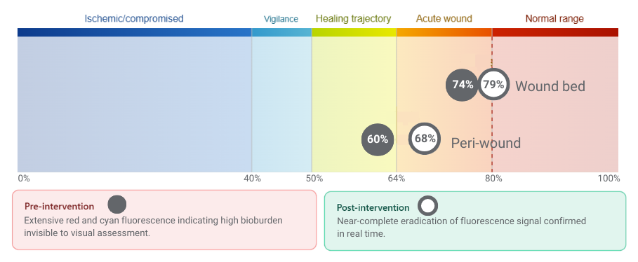
The Impact of the snapshot suite
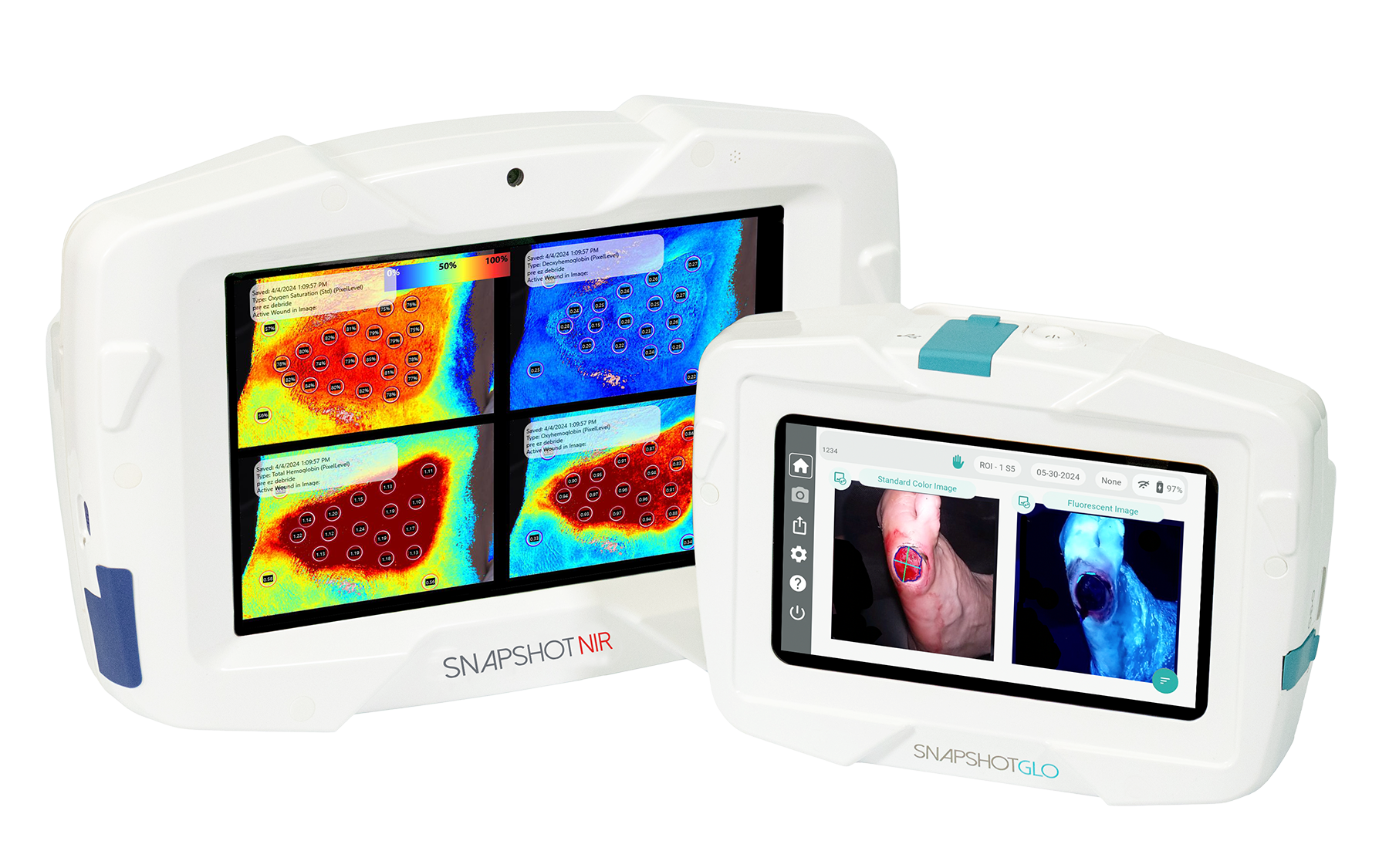
“Without the Snapshot Suite, I would have managed this wound based on what I could see, and what I could see was not telling the whole story. These devices changed the intervention and then confirmed it worked. That’s a level of certainty we haven’t had before.”