
MEDIA KIT
WHO WE ARE
Near the start of the millennium, the National Research Council of Canada had completed a lab model to show proof of concept of near-infrared spectroscopy (NIRS) to look at human tissue for the concentration of oxygen. Kent’s Founder and Executive Chairman, Don Chapman, was intrigued by its potential in the medical setting and Kent Imaging was formed. Kent Imaging was Don’s last project, as he passed in February 2025.
Initial funding from the founder and several private investors, coupled with stringent cost controls, allowed the organization to move through several years and iterations of design and field-testing programs until a smaller, sleeker, handheld unit was devised.
LEADERSHIP TEAM
Pierre Lemire CEO
Chris Tiemann VP, Global Sales
Sandra Jenks VP, Global Marketing
Chamith Rathnayake VP, Manufacturing Operations
Christine Shettel VP, Clinical Services
Jason Nessler VP, Product Development
Dr. Jeffrey Niezgoda CMO, Limb Preservation
Dr. Glyn Jones CMO, Surgery
Karel Zuzak Senior Medical Scientist
Jennie Feight VP, Health Policy
Hitalo Arume VP, Regulatory Affairs and Quality Assurance

Company Milestones
June 2026 SnapshotNIR software update to include full integration with Epic EHR PRESS RELEASE
May 2026 Expansion into Hong Kong and Macau, and the GCC and Iraq PRESS RELEASE
February 2026 Celebrates 20 year anniversary PRESS RELEASE
December 2025 Health Canada approval for SnapshotNIR KD205 PRESS RELEASE
September 2025 Launches SnapshotGLO: A New Diagnostic Imaging Solution for Bioburden Detection PRESS RELEASE
SnapshotNIR Receives CE Marking: Expansion into the European Union and United Kingdom PRESS RELEASE
April 2025 Software update to SnapshotNIR to include a DocNow EHR integration, and WoundExpert EHR web-based compatibility and updated report templates to include hemoglobin view images PRESS RELEASE
2024 Advancement to SnapshotNIR (KD205) with an algorithm update that optimizes NIRS imaging for all areas of the body, in nearly all skin tones, effectively addressing the melanin barrier in medical imaging PRESS RELEASE
2023 Expanded sales to new international markets including, Israel and Egypt.
2022 Snapshot V4.0 (KD204T) was released with advanced wound measurement functions and customizable reporting.
2017 The FDA cleared Snapshot V2.0 (KD203), the next iteration of the NIR imaging device, featuring a battery-powered handheld footprint with no docking station or wires.
2015 Pierre Lemire was welcomed to the team as an experienced CEO to lead the development of medical technology solutions and manage the company’s expansion into the future.
Changing the way clinicians treat acute and chronic wounds with actionable Tissue insights they never had before



The Snapshot Suite
Two invisible problems. One device pairing that sees both.
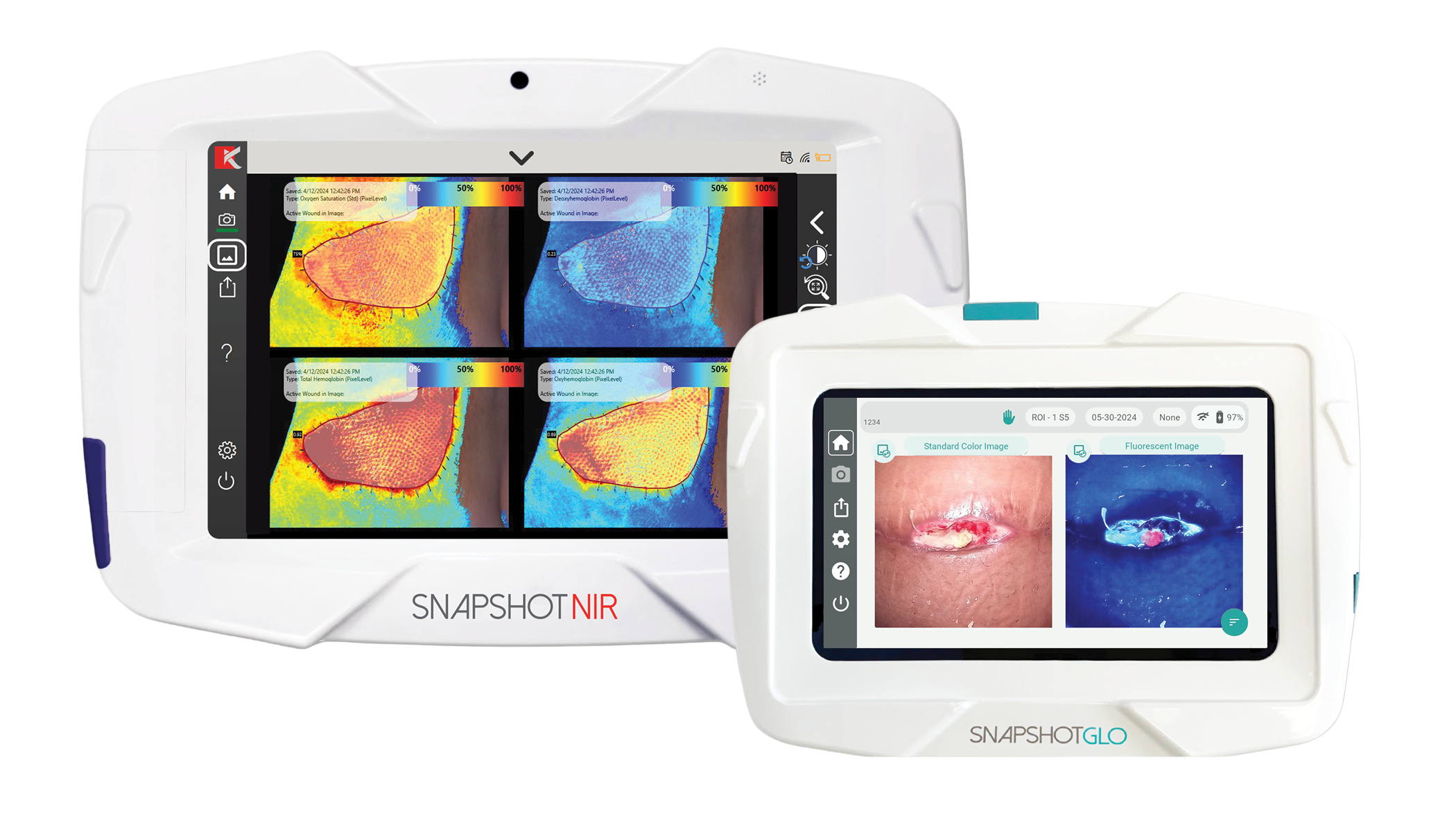
The Snapshot Suite combines two complementary imaging technologies to provide clinicians with objective, real-time physiologic data at the bedside. Used together, SnapshotGLO and SnapshotNIR can help detect the barriers of wound healing (bacteria & hypoxia), direct targeted care (clean here, debride here, offload here), and support clinical decisions with visual proof and documented outcomes.
Understanding the capacity of a wound to heal is essential across health fields including chronic wounds, surgical cases and vascular diseases.
14-24% of patients with diabetes mellitus will develop a diabetic foot ulcer during their lifetime and up to 1 in 4 of those patients will undergo an amputation - that’s an amputation every 30 seconds annually (1, 2).
Approximately 50-60% of diabetic foot ulcers (DFUs) become infected (3).
There is a 46% risk of death within the first 5 years after a minor amputation and 57% risk after a major amputation (4).
In reconstructive surgery, 1 in 3 mastectomies result in post-operative complications over the next two years and almost 20% of patients will require further surgery (5).
In vascular disease assessments like Peripheral Arterial Disease and Chronic Limb-Threatening Ischemia, ‘gold standard’ diagnostics, ABIs, will miss the diagnosis up to 60% of the time (6).

The innovative SNAPSHOTNIR
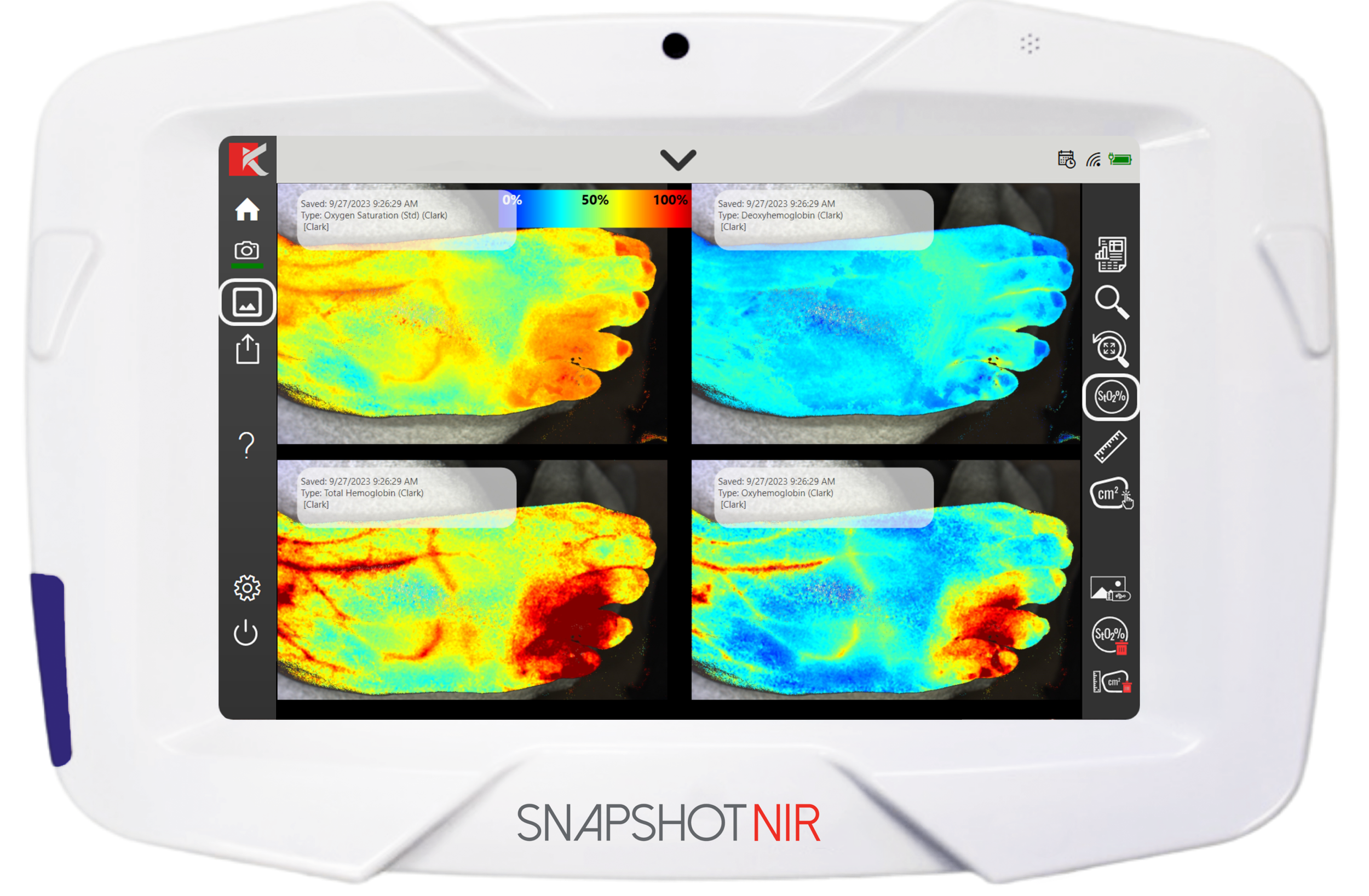
SnapshotNIR is a FDA-cleared, portable imaging device that uses near-infrared spectroscopy (NIRS) to non-invasively measure the content of oxygen bound to hemoglobin at the tissue/cellular interface (StO2), just below the surface of the tissue being imaged.
SnapshotNIR directly addresses the need for objective, real‑time assessment of tissue viability at the point of care. With a click of a button on the non-invasive, handheld device, a StO2 oxygenation map is produced of the wound bed and peri-wound, or general region of interest.
Clinicians can view oxyhemoglobin, deoxyhemoglobin, and total hemoglobin trends which can support assessment of underlying conditions or tissue healing capacity (7). These parameters help clinicians evaluate tissue viability, healing potential, and underlying physiological contributors such as ischemia, venous congestion, or infection risk.

Advancing healthcare
Kent’s proprietary algorithm expands the technology use to include patients with high melanin content. PRESS RELEASE

Intra-operative surgery
SnapshotNIR can predict skin flap necrosis during post-mastectomy breast reconstructions reducing complication rates and improving patient quality of life. PUBLICATION

Largest dataset study using SnapshotNIR
Real-world evidence study uses SnapshotNIR on 4,000+ wounds to deliver targeted care and improve wound healing outcomes. PUBLICATION

Reduce physician burden
Doctors need more time with patients and less time in paperwork. Quick, accurate, repeatable objective data and ease-of-use allows physicians to take control of their time without compromising on patient care. REIMBURSEMENT

Peripheral Arterial Disease (PAD) novel protocols
SnapshotNIR has been used as the technology for two peer-reviewed, published protocols for evaluating PAD: The Plantar-Palmar Index and the Provocative Leg Manuever, updating the traditional assessment methods.
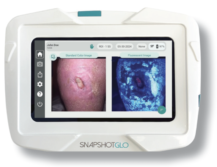
SnapshotGLO is a FDA-cleared, non-invasive, handheld imaging device that uses bacterial autofluorescence imaging (BAFI) technology. By emitting specific light wavelengths to excite the natural fluorescence of bacteria, SnapshotGLO detects high bacterial loads >10⁴CFU/g, pinpointing harmful bacteria that are otherwise invisible to the naked eye.
BAFI provides real-time bacterial detection at the point of care with 87-100% accuracy, performing 3-4x better than standard of care alone (8). BAFI uses ultraviolet light to excite endogenous bacterial fluorophores supporting real-time mapping of harmful bacteria at the bedside (9).
The glow-up wound care deserves: SnapshotGLO


Detect distinct bacterial strains
Support targeted treatments with high-resolution autofluorescence imaging detecting cyan and red fluorescence.
Objective data at the bedside
Efficient imaging workflows with SnapshotGLO help clinicians detect bacteria bioburden and aid in targeted treatments and support antibiotic stewardship.

Tracking wound healing progress
Digital wound measurement and non-invasive serial imaging allow clinicians to track and document healing progress strengthening clinical decisions and documentation for reimbursement.
Customer Success
-
![Misael alonso, MD, headshot near hyperbaric chambers.]()
Misael Alonso
MD, FACP, CWSP, FAPWCA
“With SnapshotNIR, I can take a picture and get a result within mere seconds.”
-
![Tyler Sexton standing next to the SnapshotNIR device]()
Tyler Sexton
MD, MAPWCA
“Without the Snapshot Suite, I would have managed wounds based on what I could see, and what I could see was not telling the whole story. These devices changed the intervention and then confirmed it worked. That’s a level of certainty we haven’t had before.” -
![Dr. Naz Wahab and Dr. Michael Kooyman holding SnapshotNIR device]()
Naz Wahab
MD, CEO Wound Care Experts Speciality
“Snapshot gives us a better idea of what’s actually happening in the wound, underneath the skin.” -
![Martha and amanda in wound care plus uniforms]()
Martha R. Kelso
RN, CHWS, DAPWCA, HBOT, Chief Executive Officer for Wound Care Plus and Head of Research for Clinical Outcomes and Research Alliance (CORA)
“SnapshotNIR lets us look below the skin’s surface, beyond what we can see with the naked eye. It changes the game.”



Media features
-

Global News Gamechangers
-

Disruption Magazine
-

Worldwide Business with Kathy Ireland
-

Executive Global
-

Health Tech Magazine
-

Executive Global

in the hands of clinicians worldwide
PRESS AND MEDIA CONTACT:
marketing@kentimaging.com
Kent Imaging Boilerplate
About Kent Imaging
Based in Calgary, Alberta, Canada, Kent Imaging is a leading innovator in advanced diagnostic imaging technology. The company designs, manufactures, and markets medical devices that support real-time decision-making in wound care, vascular, and surgical specialties. Through patented technologies, Kent continues to deliver groundbreaking solutions that support healthcare systems around the world.
The Snapshot product suite now includes two powerful imaging devices: SnapshotNIR, a near-infrared tissue oxygenation imaging device initially cleared by the FDA and Health Canada in 2017 and CE-marked in 2025; and SnapshotGLO, a bacterial autofluorescence imaging device that received FDA 510(k) clearance in 2025. SnapshotNIR is supported by clinical evidence demonstrating its role in enhancing clinical decision-making, while SnapshotGLO provides real-time insight into wound bioburden presence and distribution. Together, the Snapshot product family strengthens clinicians' ability to detect, direct, and protect—promoting treatment consistency, supporting improved workflow, and contributing to better patient care worldwide.
SnapshotGLO® is a registered trademark of Kent Imaging Inc.
SnapshotNIR® is a registered trademark of Kent Imaging Inc.
References:
Sen CK. Human wound and its burden: updated 2022 compendium of estimates. Advances in wound care. 2023 Dec 1;12(12):657-70.
Dayya D, O'Neill OJ, Huedo-Medina TB, Habib N, Moore J, Iyer K. Debridement of diabetic foot ulcers. Advances in wound care. 2022 Dec 1;11(12):666-86.
Armstrong, D. G., Tan, T.-W., Boulton, A. J. M., et al. (2023). Diabetic foot ulcers: A review. JAMA, 330(1), 62–75.
Armstrong DG, Swerdlow MA, Armstrong AA, Conte MS, Padula WV, Bus SA. Five year mortality and direct costs of care for people with diabetic foot complications are comparable to cancer. Journal of foot and ankle research. 2020 Dec;13:1-4.
Bennett KG, Qi J, Kim HM, Hamill JB, Pusic AL, Wilkins EG. Comparison of 2-Year Complication Rates Among Common Techniques for Postmastectomy Breast Reconstruction. JAMA Surg. 2018;153(10):901–908. doi:10.1001/jamasurg.2018.1687.
AbuRahma AF, Adams E, AbuRahma J, Mata LA, Dean LS, Caron C, Sloan J. Critical analysis and limitations of resting ankle-brachial index in the diagnosis of symptomatic peripheral arterial disease patients and the role of diabetes mellitus and chronic kidney disease. Journal of vascular surgery. 2020 Mar 1;71(3):937-45.
Oropallo, A., Ortega-Loayza, A. G., Korzendorfer, H., James, F., Dotson, P., Khimchenko, A., ... & Sonenblum, S. E. (2025). Advancing chronic wound care with near-infrared spectroscopy imaging: clinical applications, measurement parameters, and insights into healing dynamics. Wounds: a compendium of clinical research and practice, 37(10), 384-392.
Oropallo, A. R., Andersen, C., Abdo, R., Hurlow, J., Kelso, M., Melin, M., & Serena, T. E. (2021). Guidelines for point-of-care fluorescence imaging for detection of wound bacterial burden based on Delphi consensus. Diagnostics, 11(7), 1219. https://doi.org/10.3390/diagnostics11071219.
Badrie, S., Moore, Z., Patton, D., O'Connor, T., & Derwin, R. (2025). The clinical utility of autofluorescence imaging for bacterial detection in wounds: a systematic review. International wound journal, 22(6), e70678.