
Improving Debridement Precision and Wound Bed Preparation Using SnapshotGLO
with Dr. Tyler Sexton, MD, MAPWCA
Clinical Case Series: Real-World Applications
Case 1: Navigating Patient Compliance & Bioburden Management
A patient with a chronic venous leg ulcer (VLU), complicated by venous insufficiency and fragile skin.
Case 2: Confirming Debridement Success in a Complex Diabetic Ulcer
A patient with a large, chronic, lower extremity ulcer presenting with bilateral edema, hyperkeratosis, and diabetes.
The invisible enemy
Chronic wounds that have stalled in their healing trajectory require more than just a “problem-solving mindset”—they require actionable, real-time data. Early detection and management of bacterial burden can reduce the risk of infection and increase the likelihood of wound closure (1). Visual and physical assessment often fail to identify high bacterial loads, leading to ‘blind’ debridement. This risks leaving behind harmful bacteria that fuels chronicity, or potentially may result in removing viable tissue.
82% of wounds with high bacterial loads (>10⁴CFU/g) show no clinical signs of infection (2)
Up to 50% of patients may not present with signs and symptoms of a systemic infection (3)
60% of all diabetic foot ulcers may become infected (4)

SnapshotGLO device.
the GLO Effect
Bacterial autofluorescence provides real-time bacterial detection at the point of care with 87–100% accuracy, performing 3-4x better than standard of care alone (5). By emitting specific light wavelengths to excite the natural fluorescence of bacteria, SnapshotGLO detects high bacterial loads >10⁴CFU/g (6), pinpointing harmful bacteria that are otherwise invisible to the naked eye.
Objective Evidence: Move beyond gut feeling to clearly visualize medical necessity and support data-driven debridement decisions.
Efficiency: Reduce time spent on ineffective or incomplete interventions.
Quality: Assess for the need of enhanced debridement for adequate wound bed preparation.
Education: Increase patient buy-in by showing them the “invisible” bacteria on their wound.
Documentation: Document procedural success with pre- and post-intervention images providing medical necessity and clinical outcome.
The Result: Precise, conservative, and patient-centered debridement that effectively re-starts the acute healing process, ensuring intervention efficacy.
Clinical Case Series: Real-World Applications
Navigating Patient Compliance & Bioburden Management
A patient with a chronic venous leg ulcer (VLU), complicated by venous insufficiency and fragile skin.
Pre-intervention: Initial imaging with SnapshotGLO revealed significant cyan fluorescence, indicating a bacterial load of >10⁴CFU/g. This objective data prompted the clinician to obtain a culture and proceed with targeted debridement.
Post-intervention: Imaging with SnapshotGLO demonstrated a decrease in harmful bacteria, but not eradication. The patient complained of pain during the debridement and did not want to continue with further treatment.
Clinical Utility: The clinician used real-time images to educate the patient on the presence of remaining bacteria. This visual evidence served as a powerful tool for patient education, illustrating why subsequent treatments would be optimal: to prevent infection and promote healing.
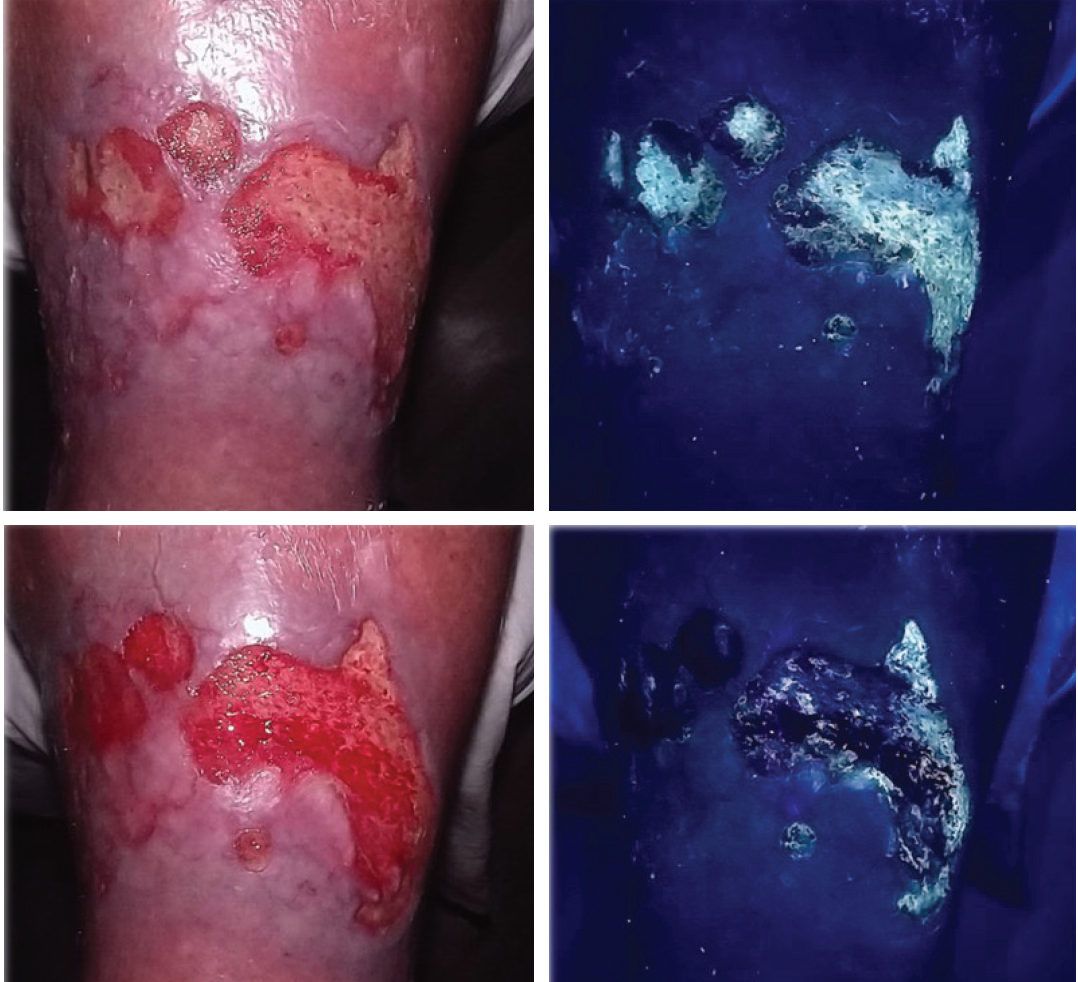
Image 1. Venous leg ulcer. SnapshotGLO images. Pre-debridement (top 2 images) clinical and bacterial autofluorescence images. Post-debridement (bottom 2 images) clinical and bacterial autofluorescence images.
Confirming Debridement Success in a Complex Diabetic Ulcer
A patient with a large, chronic, lower extremity ulcer presenting with bilateral edema, hyperkeratosis, and diabetes. Given the wound’s chronicity and presentation (erythema, slough, odor, and maceration), high bacterial burden was suspected but not visually quantifiable via standard clinical assessment.
Pre-intervention: SnapshotGLO confirmed the suspicion, pinpointing specific areas of high bacterial loads as cyan fluorescence across the wound bed. Armed with objective data, the clinician performed ultrasonic debridement using a qoustic wound therapy device (Arobella) to remove devitalized tissue and bioburden.
Post-intervention: Imaging confirmed the total eradication of cyan fluorescence, leaving a clean, granular, bleeding base.
Clinical Utility: The device provided data to support the clinical decisions and interventions performed by the provider, confirming that the debridement was successful. The patient tolerated the the ultrasonic debridement well, and the wound demonstrated reduced odor and immediate improvement in appearance. The wound was now adequately prepared for the next phase of healing. This provided the clinician with objective data that demonstrated that the wound bed was optimally prepared for advanced therapies, moving the wound from a stalled inflammatory state into the proliferative phase.
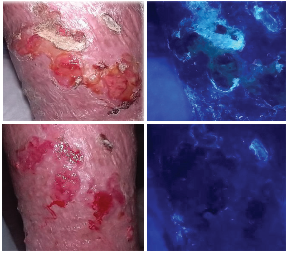
Image 2. Chronic lower limb ulcer. SnapshotGLO images. Pre-debridement (top 2 images) clinical and bacterial autofluorescence images. Post-debridement (bottom 2 images) clinical and bacterial autofluorescence images.

Dr. Tyler Sexton, MD
“SnapshotGLO imaging provided objective confirmation of bioburden, validated the effectiveness of debridement, and offered measurable evidence of tissue status.”— Dr. Tyler Sexton
Dr. Tyler Sexton, MD, MAPWCA, is the Chief Medical Officer (CMO) at Brevard Regional Hyperbaric Center, Melbourne, Florida; CMO, UrgentFlex Orlando FL; CMO, True Hyperbar Rx, Orlando, FL; CMO, Coastal HyperbaRx Ocean Springs, MS; and CMO, Precision Hyperbarics, Tampa, FL.
More SnapshotGLO resources:
Check out the clinical posters
See the applications of the device
References:
Li, S., Renick, P., Senkowsky, J., Nair, A., & Tang, L. (2021). Diagnostics for wound infections. Advances in Wound Care, 10(6), 317–327. https://doi.org/10.1089/wound.2020.1216
Le, L., et al. (2021). Diagnostic accuracy of point-of-care fluorescence imaging for the detection of bacterial burden in wounds: Results from the 350-patient fluorescence imaging assessment and guidance trial. Advances in Wound Care, 10(3), 123–136. https://doi.org/10.1089/wound.2020.1307
Ding, X., Tang, Q., Xu, Z., et al (2022). Challenges and innovations in treating chronic and acute wound infections: From basic science to clinical practice. Burns & Trauma, 10, tkac014. https://doi.org/10.1093/burnst/tkac014
Armstrong, D. G., Tan, T., Boulton, A. J. M., & Bus, S. A. (2023). Diabetic foot ulcers: A review. JAMA, 330(1), 62–75. https://doi.org/10.1001/jama.2023.10578
Oropallo, A. R., Andersen, C., Abdo, R., Hurlow, J., Kelso, M., Melin, M., & Serena, T. E. (2021). Guidelines for point-of-care fl uorescence imaging for detection of wound bacterial burden based on Delphi consensus. Diagnostics, 11(7), 1219. https://doi.org/10.3390/diagnostics11071219
SnapshotGLO (KB100) FDA 510(k) K242669
Thank you to Dr. Tyler Sexton, MD and his clinical team for these case examples.
Dr. Tyler Sexton is the Chief Medical Officer (CMO) at Brevard Regional Hyperbaric Center Melbourne Florida; CMO, UrgentFlex Orlando Fl; CMO, TrueHyperbarRx , Orlando, Fl; CMO, Coastal Hyperbarx Ocean Springs, MS; and CMO, Precision Hyperbarics, Tampa, Fl.