
SnapshotNIR guides HBOT treatment for a necrotic facial wound leading to full wound closure
Case courtesy of Tyler Sexton, MD, MAPWCA
42% to 82% tissue oxygen saturation, from necrotic tissue, to healed wound, no surgery needed.
What if you could see tissue healing in real time — and use that data to avoid surgery altogether?
In this compelling case study, Dr. Tyler Sexton shares how SnapshotNIR guided the treatment of a complex necrotic facial wound caused by osteoradionecrosis. Using near-infrared imaging to monitor tissue oxygen saturation, his team was able to track the impact of hyperbaric oxygen therapy session by session — watching StO2 levels rise from ~42% at baseline to ~82% after 17 sessions.
The result? Full wound closure. No surgery. A better quality of life for the patient.
Download the full case study to see how objective imaging data is transforming wound care decisions.
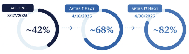
Image 1 Serial imaging with SnapshotNIR from baseline to 17 HBO dives showed a progressive increase in StO2 supporting clinical decisions and patient adherence to the treatment plan.
The Impact of SnapshotNIR
“SnapshotNIR findings validated our clinical judgment regarding impaired perfusion but also revealed more detailed, localized deficits than what were appreciated on visual inspection alone. It confirmed the suspected need for adjunctive oxygenation and allowed for more precise wound mapping, reinforcing the use of HBOT. It added a quantifiable dimension to ongoing wound surveillance not achievable through visual exam alone.”
