
The Colors of Wound Care - What Does the Snapshot Suite Expose in Wound Care?
A new patient arrives at your clinic and they have a persistent wound that has not shown healing progression within four weeks despite implementation of standard of care and no signs of infection.
As a clinician, when you are assessing a chronic wound like this, you’re looking for the subtle shifts in tissue health that dictate your next move. However, even the most seasoned experts are limited by what the human eye can perceive.
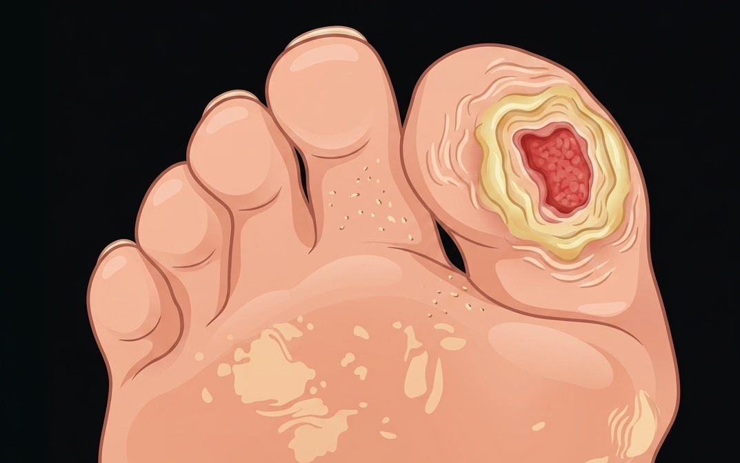
Image 1: Wound on great toe, depicting a wound seen only through visual assessment (AI generated image).
Two of the most critical questions in chronic wound care are:
"Is there adequate oxygenation to support healing?"
"Is there bacterial bioburden stalling progress?"
These questions often remain unanswered during a standard visual exam. You can’t see the microcirculation on visual inspection. 82% of wounds with high bacterial load don’t show clinical signs and symptoms of infection (1). Relying on this visual and physical assessment leaves too much uncertainty on the table.
The Snapshot Suite (SnapshotNIR and SnapshotGLO) helps you move into the 21st century with your assessment. These real-time, bedside imaging technologies look "below the surface" and expose factors that affect wound healing: poor tissue oxygenation and harmful levels of bacteria.
The colors of tissue oxygenation: SnapshotNIR
Oxygen is fuel for cellular repair (2). The visual appearance of tissue doesn't always reflect the metabolic reality beneath the surface. The patient whose wound has stalled after 4 weeks may have diagnosed vascular disease impacting the healing process.
SnapshotNIR is a non-contact, near-infrared (NIR) technology that measures tissue oxygen saturation (StO2), oxyhemoglobin (HbO), deoxyhemoglobin (Hb), and total hemoglobin (TotHb) across nearly all skin tones. NIR light harmlessly passes through superficial tissue to interact with hemoglobin in the microvasculature. The light is either absorbed or reflected back based on the oxygen content of the hemoglobin. The device processes the reflected light to create a color-coded map of high or low areas of oxygenation in both the wound bed and peri wound area. SnapshotNIR data allows clinicians to visualize tissue viability in real-time, enabling more confident clinical decision-making.
Decoding the Colors of SnapshotNIR
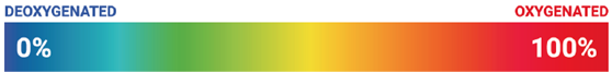
Cooler hues (Blues, Greens): These can present as low StO2 and high deoxyhemoglobin values, indicating poor tissue oxygenation. This may indicate underlying vascular conditions such as venous or arterial insufficiency and support the clinical decision to refer the patient for a vascular assessment (2).
Warmer hues (Oranges, Reds): These can present as higher StO2 and increased oxyhemoglobin values, indicating adequate tissue oxygenation. In a chronic wound, an increase in StO2 and warmer tissue colors may indicate a healing trajectory (2).
Serial imaging can monitor the trends of hemoglobin over time, indicating that a wound is trending towards closure as the oxyhemoglobin concentrations increase and the deoxyhemoglobin concentrations decrease.
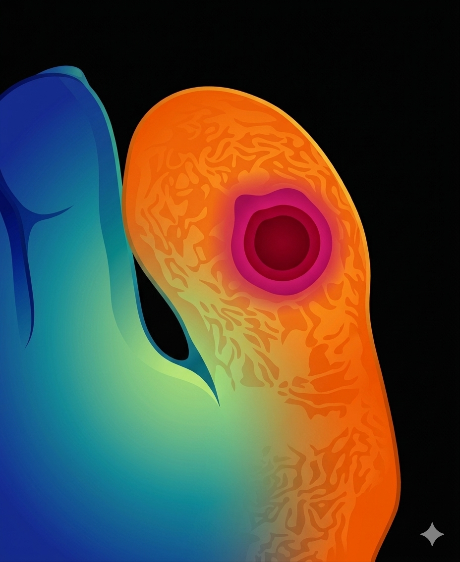
Image 2: Wound on great toe, seen through the lens of SnapshotNIR with warmer hues and cooler hues depicting the general colors of tissue oxygenation (AI generated image).
The glow of fluorescing bacteria: SnapshotGLO
Waiting for the obvious signs and symptoms of wound infection, that may never appear, is a missed opportunity to intervene before complications impact your patient’s progress (1). The patient whose wound has stalled at 4 weeks may have harmful bacteria impeding the healing process.
SnapshotGLO is a portable, handheld bacterial autofluorescence imaging device that can detect high bacterial loads of >10^4 CFU/g, in real-time. The device uses concentrated ultraviolet (UV) light to excite the natural fluorescent properties of most bacteria. Instead of guessing based on unreliable clinical signs and symptoms, you see the bacteria as a "glow" on the screen.
Decoding the Colors of SnapshotGLO
Cyan Fluorescence: This can indicate the presence of pyoverdine, a siderophore specifically produced by Pseudomonas aeruginosa.
Red Fluorescence: This can indicate porphyrin siderophore, often emitted by many bacteria during iron consumption, and is a common infection biomarker.
Detecting elevated bacterial load can assist clinicians in performing more effective cleansing techniques, more targeted debridement interventions, and more precise tissue cultures when necessary (3).
Image 3: Wound on great toe, seen through the lens of SnapshotGLO depicting the general glowing colors of fluorescing bacteria in cyan and red fluorescence (AI generated image).
Seeing the colors of wound care with the Snapshot Suite
It is difficult to fight what you cannot see. Relying on only your basic senses is a traditional approach to a modern clinical challenge. The wound that looked clean on week six might be losing the battle beneath the surface. By integrating the Snapshot Suite, you can bridge the gap between clinical intuition and objective data. Whether it’s visualizing the oxygen map with SnapshotNIR or uncovering the bacterial glow with SnapshotGLO, you are no longer just seeing the surface; you are treating the underlying wound.


References
Le, Lam, et al. "Diagnostic accuracy of point-of-care fluorescence imaging for the detection of bacterial burden in wounds: results from the 350-patient fluorescence imaging assessment and guidance trial." Advances in Wound Care 10.3 (2021): 123-136.
Oropallo, Alisha, et al. "Advancing chronic wound care with near-infrared spectroscopy imaging: clinical applications, measurement parameters, and insights into healing dynamics." Wounds: a compendium of clinical research and practice 37.10 (2025): 384-392.
Oropallo, A. R., Andersen, C., Abdo, R., Hurlow, J., Kelso, M., Melin, M., & Serena, T. E. (2021). Guidelines for point-of-care fluorescence imaging for detection of wound bacterial burden based on Delphi consensus. Diagnostics, 11(7), 1219. https://doi.org/10.3390/diagnostics11071219