
Non-invasive measurement of tissue oxygenation through Near-Infrared Spectroscopy
The problem: Assessment of Chronic Wounds
When it comes to chronic wound management, acute wounds in reconstructive surgery, or limb preservation, oxygenation of the tissue is vital. Tissue oxygenation refers to oxygen saturation which is a measure of the percentage of dissolved oxygen in tissue. Tissue oxygenation takes place when oxygen molecules enter the tissues of humans when blood is oxygenated in the lungs through the oxygen molecules traveling from the air and into the blood.
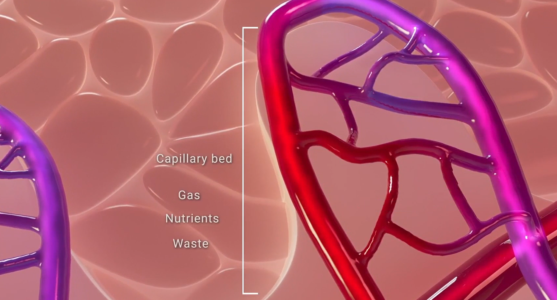
Gas exchange at the capillary level in the microvasculature.
The growing prevalence of chronic and non‑healing wounds, driven largely by an aging population and rising comorbidities, continues to place a significant burden on healthcare systems (Sen, 2023). Conditions such as diabetes, peripheral arterial disease (PAD), and neuropathy impair the body’s ability to heal chronic wounds (Gornik et al., 2024), increasing the risk of stalled healing, recurrence, infection, amputation, and premature mortality (Armstrong et al., 2025; Armstrong et al., 2020).
Patients are at risk of higher complication rates when presenting with compromised vascular status and comorbidities including flap surgeries and oncologic surgeries such as post-mastectomy breast reconstruction (Hill et al., 2024; Moritz et al., 2023).
Across both wound care and surgical care, tissue hypoxia and ischemia remain among the strongest predictors of non‑healing wounds, surgical dehiscence, flap failure, and necrosis (Oropallo et al., 2025; Hill et al., 2024). Adequate oxygenation is essential for every phase of tissue repair (Oropallo et al., 2025).
Current Assessment Methods
There is a gap in the medical system for fast, accurate, repeatable technology that is cost-effective and efficient. The current gold standard approach of clinical monitoring in wound care is:
Visual inspection
Physical measurements with a paper ruler
Vascular assessments to determine oxygenation
While commonly used, these approaches provide limited insight into microcirculatory health (Geskin et al., 2022), the level at which healing is most critical.
Clinicians turn to conventional tests for vascular assessment of blood and tissue viability, including the Ankle-Brachial Index (ABI), Toe-Brachial Index (TBI), and Transcutaneous Oxygen Pressure (TcPO2). These methods each have major disadvantages compared to near-infrared spectroscopy (NIRS) imaging (Oropallo et al., 2025).

Ankle-brachial index assessment (Chaudru et al., 2016)

Transcutaneous oximetry (López-Moral et al., 2023)
An innovative solution for wound assessment: near-infrared spectroscopy
As an alternative to direct O2-sensing approaches, near-infrared spectroscopy (NIRS) has demonstrated the ability to accurately quantify tissue oxygen saturation levels with zero patient contact. NIRS is an optical method consisting of illuminating chemical compounds that absorb, reflect, and scatter light directed at that compound. Faster detection of critical changes in tissue oxygenation could enable earlier and more successful intervention when tissue complications arise. NIR light is very useful in detecting oxygenated and deoxygenated blood, which conveys a comprehensive picture of tissue health and the healing capacity of acute and chronic wounds.
snapshotnir: the Portable NIRS device
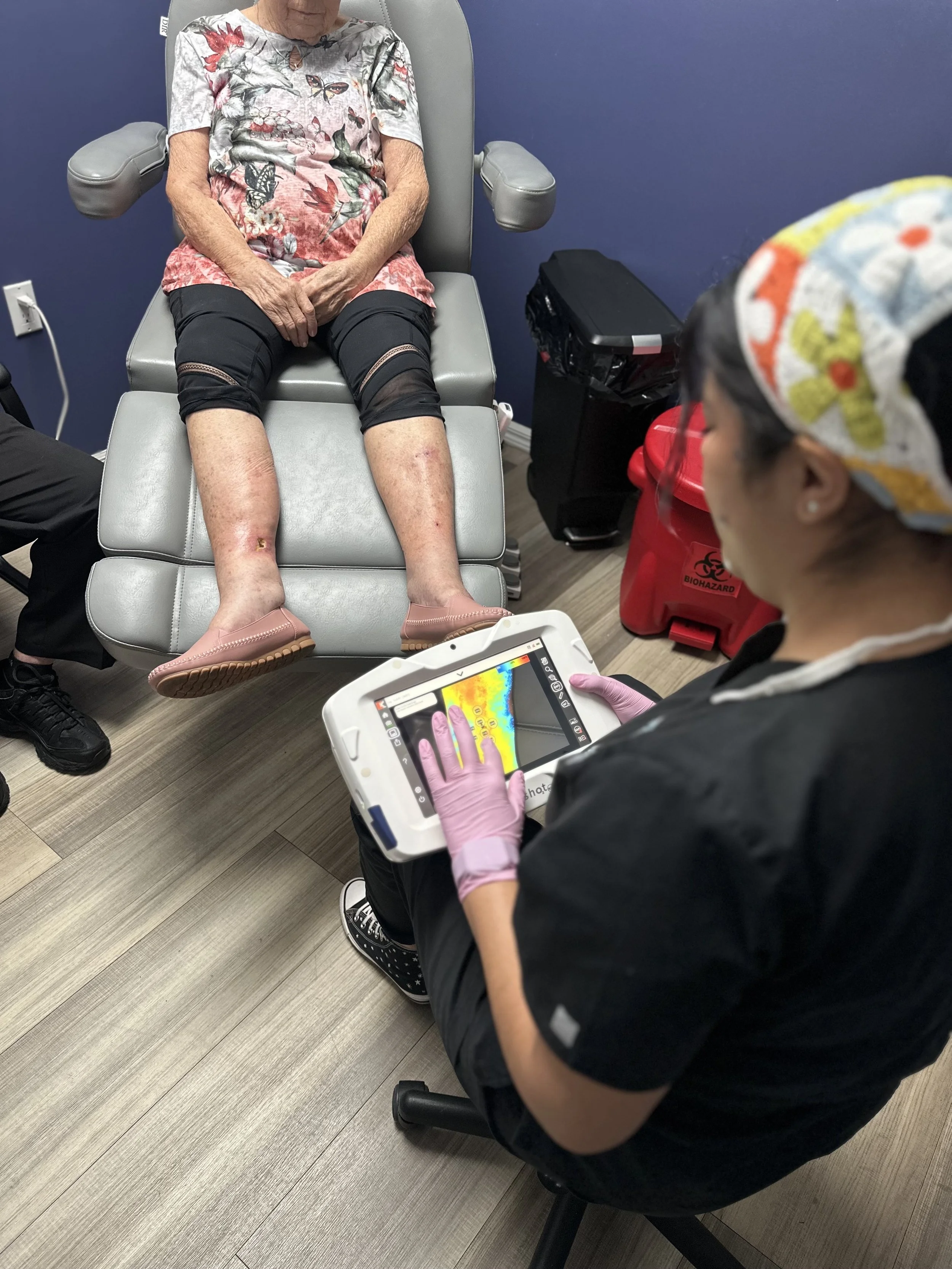
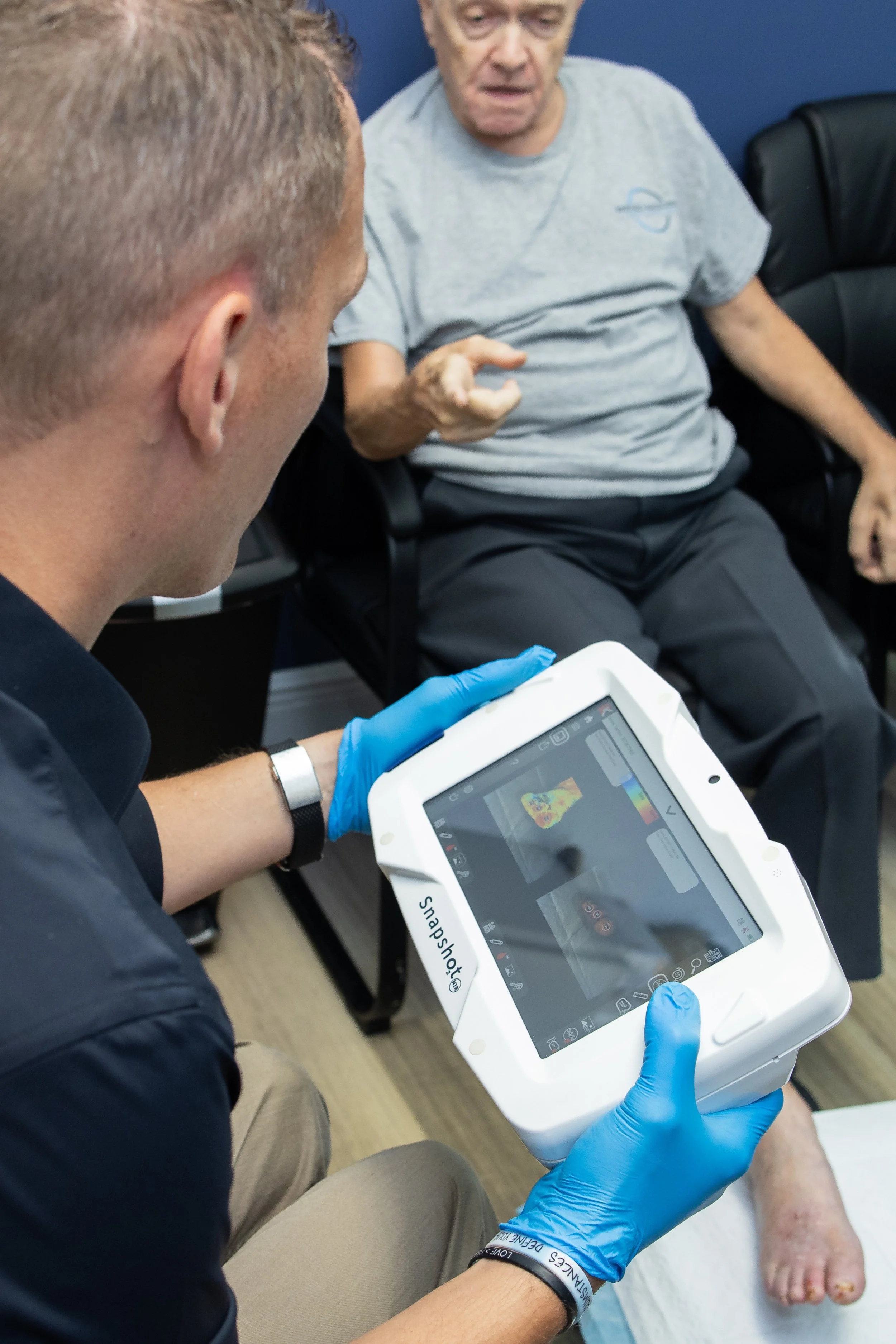
SnapshotNIR, by Kent Imaging, is a near-infrared (NIR), reflectance-based technology that measures tissue oxygen saturation in superficial tissue at the critical macrovascular level. Using multiple wavelengths of NIR light, SnapshotNIR measures the relative amounts of oxygenated and deoxygenated hemoglobin in the areas where oxygen exchange is happening. SnapshotNIR provides users with a tissue oxygenation map that can be used for medical decision-making, such as tracking and trending oxygenation, and to evaluate tissue viability.
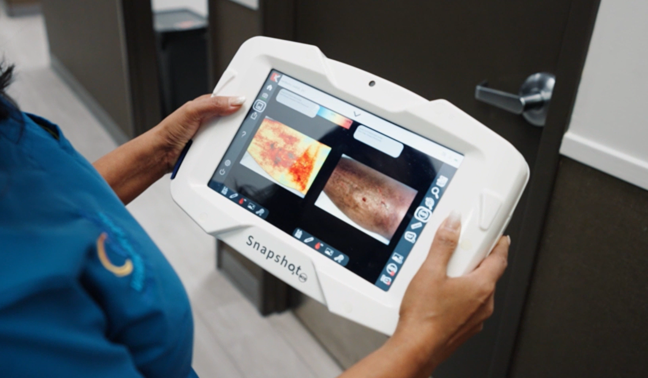
SnapshotNIR device with an image of a leg wound on the screen.
Kent Imaging has developed a cost-effective and easy-to-use point-of-care imaging device that is completely non-invasive, removing the need for patient contact or injected dyes. Snapshot NIR captures the availability of oxygenated blood in tissue in a matter of seconds, helping to provide diagnostic insight for improved decision-making throughout the continuum of care. The tracked and documented oxygenation data allows patients to be assessed for appropriate advanced wound care modalities, to monitor the therapeutic benefit, and share that information with the patient and between healthcare providers all in one handheld, portable device.
Changing your clinical practice with SnapshotNIR
Clinicians can view oxyhemoglobin and deoxyhemoglobin trends which can support assessment of underlying conditions or tissue healing capacity.
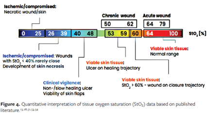
Oxygenation scale from the publication Oropallo et al., 2025.
Low oxyhemoglobin and high deoxyhemoglobin may indicate underlying venous disease or other vascular compromise hindering the wound’s ability to heal (Oropallo et al., 2025)
Very high areas of total hemoglobin can indicate inflammation, infection, or a tunneling wound (Oropallo et al., 2025)
StO2 ranges can support clinical decision-making when StO2 is <39%, between 40-69% or >70% (Oropallo et al., 2025)
Physicians can use the visual data from SnapshotNIR to:
Communicate more effectively with their colleagues for expedited referrals
Document wound status for patient monitoring and insurance reimbursement
Support clinical decision-making for interventions
Educate their patients on their prognosis and healing capacity
Providers can communicate with their patients at the bedside in any care setting, helping them better understand the underlying issue, positively impact adherence to the prescribed therapy, and build rapport by demonstrating real-time improvements.
To speak to a product expert about Kent’s imaging technologies, contact us here.
To learn more about the applications of Kent Imaging’s SnapshotNIR, click here.
Original article written Jan 6, 2022. Updated on April 17, 2026.
References
Armstrong, N. S., Armstrong, A. A., Mills, J. L., Conte, M. S., Tan, T. W., Swanson, R. S., & Armstrong, D. G. (2025). Three‐Year Recurrence in People With Diabetic Foot Ulcers and Chronic Limb Threatening Ischemia Is Comparable to Cancer. International Wound Journal, 22(8), e70724.
Armstrong, D. G., Swerdlow, M. A., Armstrong, A. A., Conte, M. S., Padula, W. V., & Bus, S. A. (2020). Five-year mortality and direct costs of care for people with diabetic foot complications are comparable to cancer. Journal of Foot and Ankle Research, 13(1), 1-4. https://doi.org/10.1186/s13047-020-00404-z
Chaudru S, De Müllenheim PY, Le Faucheur A, Kaladji A, Jaquinandi V, Mahé G. Training to perform ankle-brachial index: systematic review and perspectives to improve teaching and learning. European Journal of Vascular and Endovascular Surgery. 2016 Feb 1;51(2):240-7.
Geskin, G., Mulock, M. D., Tomko, N. L., Dasta, A., & Gopalakrishnan, S. (2022). Effects of lower limb revascularization on the microcirculation of the foot: A retrospective cohort study. Diagnostics, 12(6), 1320. https://doi.org/10.3390/diagnostics12061320
Gornik, H, Aronow, H, Goodney, P. et al. 2024 ACC/AHA/AACVPR/APMA/ABC/SCAI/SVM/SVN/SVS/SIR/VESS Guideline for the Management of Lower Extremity Peripheral Artery Disease: A Report of the American College of Cardiology/American Heart Association Joint Committee on Clinical Practice Guidelines. JACC. 2024 Jun, 83 (24) 2497–2604. https://doi.org/10.1016/j.jacc.2024.02.013
Hill, W. F., Kinaschuk, K., & Temple-Oberle, C. (2024). Intraoperative nearinfrared spectroscopy can predict skin flap necrosis. Plastic & Reconstructive Surgery-Global Open, 12(3), e5669. https://doi.org/10.1097/GOX.0000000000005669
López-Moral M, García-Madrid M, Molines-Barroso RJ, García-Álvarez Y, Tardáguila-García A, Lázaro-Martínez JL. Analyses of transcutaneous oxygen pressure values stratified for foot angiosomes to predict diabetic foot ulcer healing. Journal of Tissue Viability. 2023 Nov 1;32(4):480-6.
Moritz, W. R., Kinaschuk, K., & Temple-Oberle, C. (2023). Point-of-care tissue oxygenation assessment with SnapshotNIR for alloplastic and autologous breast reconstruction. Plastic and Reconstructive Surgery - Global Open, 11(7), e5113.
Oropallo, A., Ortega-Loayza, A. G., Korzendorfer, H., James, F., Dotson, P., Khimchenko, A., ... & Sonenblum, S. E. (2025). Advancing chronic wound care with near-infrared spectroscopy imaging: clinical applications, measurement parameters, and insights into healing dynamics. Wounds: a compendium of clinical research and practice, 37(10), 384-392.
Sen, C. K. (2023). Human wound and its burden: Updated 2022 compendium of estimates. Advances in Wound Care, 12(12), 657-670.
https://www.sciencedirect.com/topics/biochemistry-genetics-and-molecular-biology/tissue-oxygenation
https://www.nature.com/articles/s41587-021-00866-y
https://annalsofintensivecare.springeropen.com/articles/10.1186/2110-5820-2-11