
Using Near-Infrared Spectroscopy Imaging to Manage Critical Limb Ischemia
SnapshotNIR uses NIR (Near-Infrared) light to determine tissue oxygenation saturation (StO2), one of the key indicators of tissue health. NIR light penetrates ~2-3mm into the tissue, making it ideal for microcirculation assessment, where oxygen exchange is happening. Key Publications in Wound Care covers some of the most up-to-date research on the use of Kent Imaging’s SnapshotNIR in the areas of wound care and limb preservation.
Using Near-Infrared Spectroscopy Imaging to Manage Critical Limb Ischemia. Today’s Wound Clinic®. 2019;13(9):12-15. Gopalakrishnan S, Niezgoda J, Hoffman B, Siddique S, Niezgoda JA.
Study Overview
CLINICAL DATA
Background: Critical Limb Ischemia (CLI) is characterized by prolonged ischemic rest pain, and non-healing wounds/ulcers/gangrene in one or both legs due to clinically established arterial occlusive disease. The poor prognosis of CLI demands a multidisciplinary approach involving various disciplines for rapid assessment, revascularization, and wound care management to enhance patient outcomes.
Methods: A 54-year old non-diabetic and non-smoking woman with no medication history underwent surgical intervention for left foot deformities and bunions. Postoperative assessment a week later showed advancing incisional and third digit (D3) tissue ischemia. Near-infrared imaging showed poor perfusion on D3 along the incisions and diffusely across the foot. The patient was then sent for angiography and endovascular arterial intervention to try to improve flow. Pre- and post-intervention near-infrared imaging demonstrated significant improvement in tissue oxygenation, indicating a greater influx of flow. Revascularization was followed by repeated bouts of hyperbaric oxygen therapy. This series of interventions lead to significant clinical improvement and D3 was completely healed at 18 weeks with salvage of the entire digit.

FIGURE 1. (A) Comparison of clinical presentation during 7-week follow-up and (B) 18-week follow-up. (C, D) At 18 weeks, D3 completely healed with salvage of the entire digit.
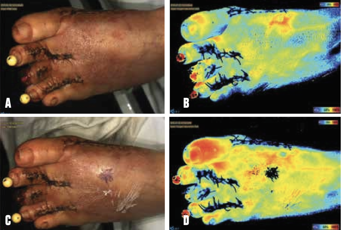
FIGURE 2. (A) Pre-angiogram clinical photograph. (B) NIRS imaging pre-angiogram. (C) Post-angiogram clinical photograph. (D) NIRS imaging post-angiogram.
Results: Using near-infrared imaging along with standard clinical practice, this patient was able to keep her foot without losing any tissue to necrosis. As a technology that focuses on perfusion and oxygenation, the near-infrared imaging device was able to reliably indicate when intervention was necessary and how successful interventions were.
Conclusion: Near-infrared imaging is a non-contact, non-invasive way of rapidly measuring tissue oxygenation and perfusion. In this case study of CLI, near-infrared imaging for longitudinal monitoring was cost-effective, time efficient, safe, and provided actionable information. The hemodynamic changes measured after endovascular revascularization and hyperbaric oxygen therapy allow clinicians to use this technology to determine the course of treatment and assess healing characteristics in CLI.
SnaphotNIR and CLTI
To hear more about how SnapshotNIR can help with patients who have chronic limb threatening ischemia, watch this webinar with Dr. Jeffrey Niezgoda, Kent Imaging CMO, and guest speaker, Dr. Craig Walker.