
Near-infrared spectroscopy in MedTech – are all devices the same?
What is NIRS in MedTech?
Near-infrared spectroscopy (NIRS) technology has been around for decades in many industries and continues to be supported by research for numerous applications.
In MedTech, NIRS has been used as a reflectance-based technology to view blood flow and perfusion underneath the skin in the macro- and microvasculature.
SnapshotNIR is a medical imaging device that provides users with tissue oxygenation maps, including tissue oxygen saturation (StO2), oxyhemoglobin, deoxyhemoglobin, and total hemoglobin. This objective data can be used for assessing therapeutic efficacy, tracking and trending wound healing, and evaluating tissue viability to help improve medical decision-making.
SnapshotNIR technology uses non-invasive wavelengths of NIR light, pointed at a region of interest on the patient, to gather data on oxygenated and deoxygenated hemoglobin within their microcirculation up to 2-4mm below the surface of the wound.

SnapshotNIR on the hemoglobin view 4-panel screen.
What makes NIRS medical imaging devices different?
All NIRS technology is not the same.
You might see differences in:
Ability of the device to accurately capture data in highly melanated skin (up to Fitzpatrick Skin Types V or VI)
Data output in various lighting conditions
Ability of the device to detect tissue and non-tissue
Features allowing the provider to complete a bedside assessment directly on the device
Data output depending on the angle of the device and position of the patient
Accuracy of tissue oxygenation readings
Different devices may have unique technical specifications and features which have the potential to impact data output. Here are four common differences that you might see when comparing NIRS MedTech.
Melanin automatic algorithm calibration
Challenge: NIRS medical imaging devices may struggle to produce objective data on patients with darker skin.
Solution: SnapshotNIR has an updated proprietary algorithm that is automatically calibrated, making it simple for clinicians to capture tissue oxygenation data on nearly all skin tones.

In this comparison, each individual had a baseline StO2 image taken with the test device and SnapshotNIR, and another image when a tourniquet was applied to a finger. With a tourniquet, blood flow should be restricted resulting in a change in StO2. You can see this change occur on the righthand side with SnapshotNIR – the tourniquet caused the finger to be more blue/green, whereas on the lefthand side, the test device did not pick up on any change with the tourniquet.
It is also apparent that the test device was unable to measure StO2 at all in the patient with higher melanin content – the resultant image does not demonstrate any reflectance-based information.
Watch the 2 min clip below to hear from Dr. Jeffrey Niezgoda and Dr. Travis Hubbuch as they demonstrate the differences in melanin correction on two NIRS medical imaging devices.
NIRS in different lighting conditions
Challenge: NIRS medical imaging devices data output could change depending on the light source in a facility, for example, LED lights.
Solution: SnapshotNIR can be used in any facility or room without clinicians needing to worry about the type of lighting at the point of care and can focus on the patient.

In this comparison, when the LED lights were on, the test device produced inaccurate results in the oximetry images, whereas SnapshotNIR was able to capture quality StO2 images regardless of the lighting conditions.
In the 1:30min clip below, Dr. Niezgoda and Dr. Hubbuch demonstrate the differences in lighting conditions on two NIRS medical imaging devices.
Tissue oxygenation saturation data
Challenge: NIRS medical imaging devices could produce data that doesn’t match the patient’s condition.
Solution: SnapshotNIR is a reliable, valid way to take serial images of a patient’s wound to assess and monitor healing.

In this comparison, the first difference is seen in the quality of the color map of tissue oxygenation in the lower leg comparison. The gradient color oxygenation from SnapshotNIR provides a clear indication of where oxygen may be reduced, compared to the test device on the lefthand side.

The second difference is in the StO2 values – the test device has a result of 6% on the plantar heel, compared to SnapshotNIR showing 66% and 75% on the two markers placed.
Watch the 2 min clip below to hear from Dr. Niezgoda and Dr. Hubbuch as they compare the tissue oxygenation images on two NIRS medical imaging devices for the lateral lower leg.
Sensitivity to detecting changes in sto2
Challenge: NIRS medical imaging devices should be able to detect tissue oxygenation and tissue perfusion when it changes from normal to low.
Solution: SnapshotNIR has the ability to place multiple markers on the patient's limb and has increased sensitivity to changes in tissue oxygenation.
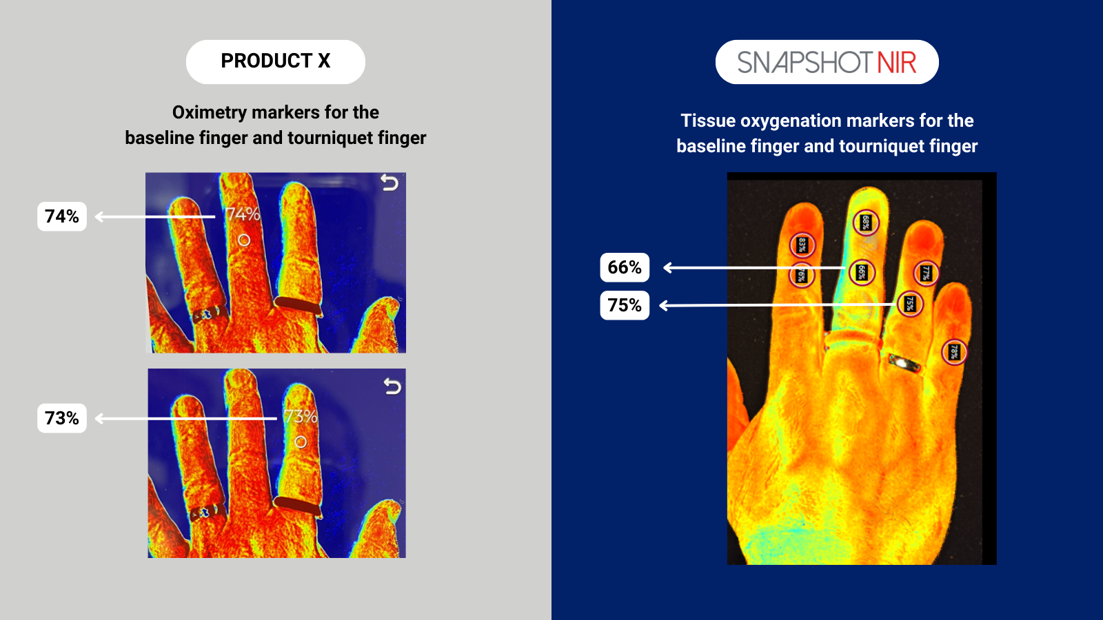
In this comparison, the test device did not show a reflective change in the baseline image (74%) to the tourniquet image (73%). Whereas SnapshotNIR markers reflected the change in tissue oxygenation from the baseline finger (75%) to the tourniquet finger (66%).
Watch the 2min clip below to hear from Dr. Niezgoda and Dr. Hubbuch as they compare the image quality and StO2 sensitivity on the devices.
Trust your medical device.
Chronic wounds are a health problem with significant reductions in quality of life and can have devastating consequences such as limb amputations and premature death. You want to trust your medical devices to assist you in making the right decision quickly.
SnapshotNIR is a diagnostic-driven, medical imaging device that can help you do that. Visualize and map tissue oxygen saturation in the capillary network, view insights into hemoglobin to make data-driven decisions and improve your patient outcomes.
Contact us and hear from a product expert to find out why you should choose SnapshotNIR.