
CPT Codes for SnapshotNIR and Documentation for Reimbursement
What CPT codes represent Near-Infrared Spectroscopy (NIRS) imaging with SnapshotNIR?
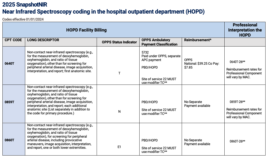
NIRS coding is captured by a primary code, an add on code, and a separate code for Peripheral Arterial Disease (PAD) screening:
0640T — primary code
Noncontact near-infrared spectroscopy (e.g., for measurement of deoxyhemoglobin, oxyhemoglobin, and ratio of tissue oxygenation), other than for screening for peripheral arterial disease, image acquisition, interpretation, and report; first anatomic site.
+ 0859T — List separately in addition to code for primary procedure, add on code
Noncontact near-infrared spectroscopy (e.g., for measurement of deoxyhemoglobin, oxyhemoglobin, and ratio of tissue oxygenation), other than for screening for peripheral arterial disease, image acquisition, interpretation, and report; each additional anatomic site (List separately in addition to code for primary procedure)
0860T — Do not report 0860T in conjunction with 0640T
Noncontact near-infrared spectroscopy (e.g., for measurement of deoxyhemoglobin, oxyhemoglobin, and ratio of tissue oxygenation), for screening for peripheral arterial disease, including provocative maneuvers, image acquisition, interpretation, and report, one or both lower extremities.
SnapshotNIR is an easy-to-use, cost-effective, and repeatable tool to assess and track the tissue oxygen level in a patient, whether at a wound site or in an area of vascular concern. Healing can be tracked and documented, with the image reports easily added to the patient record. These documented reports can be used to support medical necessity and proof of therapeutic efficacy for managed care reimbursement payments.
CPT codes, descriptions, and other data are copyright 2024 American Medical Association. All rights reserved.
CPT codes, descriptions, and other data are copyright 2024 American Medical Association. All rights reserved.
Suggested Documentation Guidelines
Documentation is critical in any profession, especially in the field of healthcare. Proper documentation is necessary to receive reimbursement from insurance payors. It is key for healthcare providers who bill for services provided to patients.
Medical necessity for any diagnostic procedure depends on one critical point: The provider must believe that the information obtained from the procedure could impact the course of treatment for the patient. An adequately documented clinical record enhances revenue cycle management. Patients get the care they desire, and the providers also get rewarded for services delivered. The following points may help to establish the validity of testing for your patients:
Indication for utilizing SnapshotNIR:
Etiology (The ‘Why’)
Appropriate diagnosis coding (may include but are not limited to)
Graft/Flap
Wound/Ulcer (i.e. Diabetic Foot Ulcer)
Pressure Injury
Burns
Traumatic
Other (i.e. autoimmune, skin tears, moisture-associated dermatitis)
Underlying factors
PAD
Diabetes with microcirculatory manifestations
Vascular disease mixed etiology (arterial and/or venous insufficiency)
Signs and Symptoms
Document any signs of compromised microcirculation/perfusion
Dusky color
Decreased temperature
Skin changes
Ulcer without improvement > two weeks
Edema
Reason for testing along with applicable underlying diagnosis (some examples listed below):
Assessment of microcirculation, oxygenation, and/or perfusion to wound and peri-wound or flap or graft site
At the start of care and subsequent visits to document trends of healing or change the plan of care
Assessment to determine the medical necessity for debridement
Adequacy of debridement to take a chronic wound from the inflammatory stage back to the acute phase
Wound bed preparation for advanced therapies
HBO Qualification
Prospective evaluation to determine if HBO is effective
If the patient requires a vascular intervention
To assess adequate wound bed preparation for advanced therapies such as cellular tissue products
Assess the need for a vascular intervention
Assess microcirculation for 4-6 weeks post-vascular reconstruction
Oxygen challenge
Description of sites (The ‘Where’)
Describe in detail the anatomical location of the wound or flap
Within each anatomical site to be imaged, note if there are multiple wounds, and if so, which ones are to be imaged
If this is a subsequent image on the same site, describe changes noted from previous images
Include a description of peri-wound tissue
Include other applicable site information (i.e., excessive edema, rubor, inflammation)
Be sure to follow general wound care, HBO and CAMPs documentation requirements
NOTE: when describing the characteristics of the site, other sections of the medical record may be referenced: “Image of L posterior lower extremity ulcer were obtained. A full description of this site may be found in the wound assessment portion of the patient record under “wound #1”.
Results and Decision-Making
Upload hemoglobin view with markers to include oxygenated and deoxygenated values to the chart
Annotate any changes or concerns (pre- and post-image)
Based on the values in hemoglobin view, describe any:
Underlying vascular disease
Trends to healing
Areas of inflammation or potential infection
Compare to previous imaging sessions as appropriate—is current therapy effective?
How will SnapshotNIR results affect your medical decision-making and plan of care?
Are additional images needed—post-intervention? Ongoing or to monitor progress?
If frequent imaging is required, why?
Referrals to appropriate specialties
Plan of care reviewed with the patient and/or caregivers
Examples:“We will send this patient for vascular consult. He/she may need surgical intervention based upon the diminished microcirculation noted in wound #1, which in my experience would not support wound healing...”
“We will begin a trial of HBO therapy for (diagnosis code), noting how the results of the imaging have shown diminished oxygenation/microcirculation, but an adequate response to an oxygen challenge examination with SnapshotNIR.”
NOTE: Reminder to link to other parts of the record as a timesaver. It is important to document fully, but referring to other the parts of the record as appropriate can prevent duplication, save time and reduce errors.
If you have further questions on reimbursement, read our FAQs page here.
If you’d like to speak to someone on our Kent Imaging Reimbursement Team, call our Toll-free Hotline 1-833-733-5368, or Email reimbursement@kentimaging.com
Disclaimer: The information provided is gathered from third party sources for informational purposes only and has not been verified with any entity responsible for coding policy. It does not represent a statement, promise or guarantee by Kent Imaging regarding coverage levels or payment for the use of SnapshotNIR. Reimbursement policies change frequently and can vary regionally and from one insurer to another. Questions regarding coverage and payment by a payor should be directed to that payor. The ultimate responsibility for documentation, coding, and claims submission lies with the physician, clinician, hospital, or other facility or provider.