
PRACTICING PREVENTION
Dr. Leon Watkins and his team at Gulf South Foot & Ankle embrace proactive podiatric care.
Last spring, when Kathryn Roth arrived at the Gulf South Foot & Ankle Clinic in Metairie, Louisiana, to have a corn on her foot trimmed, her long-time podiatrist, Dr. Leon Watkins, noticed a small ulcer on her lower left calf.
“It was about the size of a nickel,” says Dr. Watkins. “And while it didn’t look bad, she said it was actually quite painful.”
Roth, who is in her 80s, suspected the wound was due to a bug bite and told Dr. Watkins that she’d been treating it with antibiotic cream, which didn’t seem to be doing any good.
Naturally, the doctor wanted a closer look.
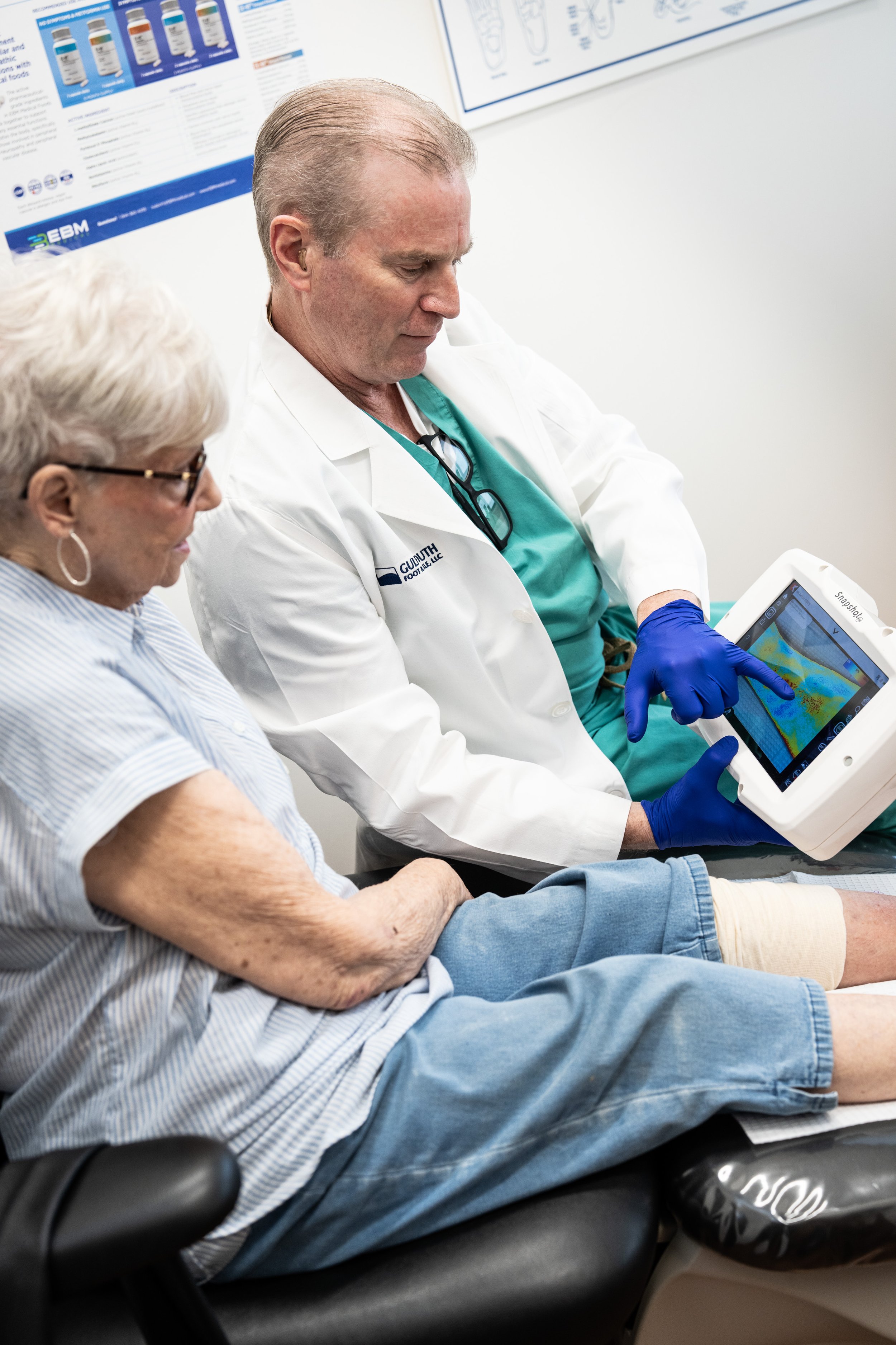
“Dr. Watkins pulled out this interesting, camera-like thing and took a picture of my leg, focusing right on the sore spot,” recalls Roth. “From that picture, he said he could see that the spot was infected inside, and I remember thinking it was so weird that such a gadget could reveal that kind of information.”
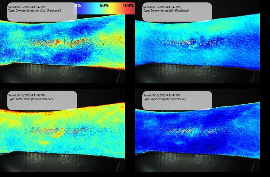
The gadget Roth is referring to is SnapshotNIR, a portable, non-invasive imaging tool that uses near-infrared light (NIR) to provide clinicians with real-time visualization of microvascular tissue oxygen saturation. Created by Kent Imaging, the diagnostic device operates much like a digital camera, capturing near-instantaneous images that allow physicians to map the ratio of oxygenated to deoxygenated hemoglobin at the wound site or in an area of vascular concern.
In Roth’s case, Snapshot images revealed that, while her wound looked okay on the outside, there was a great deal of inflammation beneath the surface.
“To the naked eye, the wound didn’t appear nearly as serious as the Snapshot images implied,” says Dr. Watkins, who, based on what he read from the device, diagnosed cellulitis and immediately sent Roth to the hospital, where she was admitted and put on IV antibiotics.
“The infection was squashed much faster thanks to SnapshotNIR,” he says. “I wouldn’t have even known there was an issue if I hadn’t used the device.”
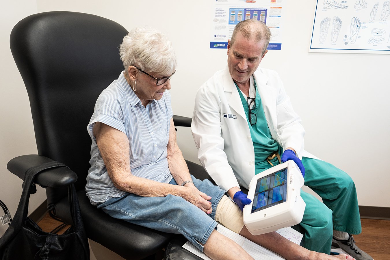
Dr. Watkins, with his patient Kathryn, reviewing images on SnapshotNIR.
LEAPING INTO ACTION
Dr. Watkins first acquired SnapshotNIR for his private podiatry clinic, Gulf South Foot & Ankle, in 2020 after seeing it in action at the New Cardiovascular Horizons conference in New Orleans. Since then, he and his colleagues utilize the device—which is shared among the clinic’s three locations—as a regular part of their practice.
“The staff love Snapshot,” he says. “We use it all the time to assess blood flow, especially in our diabetic patients.”
A large number of patients who come into Gulf South Foot & Ankle have diabetes, a disease that, according to Dr. Watkins, is quite common in the South and can lead to foot problems like chronic ulcers and vascular issues. When left untreated, these conditions can result in amputation.
As a means of preventing such outcomes, the team at Gulf South Foot & Ankle developed the Lower Extremity Amputation Prevention program, or LEAP for short. Under this voluntary, proactive initiative, diabetic patients visit the clinic once a quarter to undergo a thorough physical examination of their lower extremities, as well as x-rays, ultrasounds and other tests that vary from visit to visit.
“Because a lot of diabetics can’t feel their feet (due to peripheral neuropathy), they often don’t come to a doctor for foot ailments until they have an infection down to the bone,” says Dr. Watkins. “But with LEAP, we’ll do these x-rays and tests and find problems earlier on—problems a patient didn’t know they had. It’s been very effective in saving limbs.”
Patients in the LEAP program undergo vascular testing on their first and third quarterly visits. Before acquiring Snapshot, the doctors at Gulf South Foot & Ankle relied on basic physical examinations and ankle-brachial index (ABI) testing to assess circulation.
But now with Snapshot, says Dr. Watkins, assessing blood flow and identifying a problem is not only faster and easier, it’s much more conclusive.
“With just a regular physical exam, there’s subjectivity in diagnosing a common diabetic condition like small vessel disease in the feet,” he says, adding that he or his staff would often have to refer patients to vascular specialists for further testing and diagnosis, only to have these specialists send the patients back without aggressive intervention.
SnapshotNIR has been a game-changer in this respect. “When we use the Snapshot, diagnosis is a yes or no thing. Ultimately, it’s a much more assuring physical test that you can save and show to others, and it clearly demonstrates the degree of blood flow as well as vascular compromise,” he says.
This level of clarity has also been instrumental in enhancing patient adherence and understanding.
“Diabetics are notoriously non-compliant because they usually don’t have any pain due to neuropathy,” explains Dr. Watkins. “But when we take the Snapshots and then show them the pictures, they’re able to see that they really do have a problem. And I think it makes them take their health care more seriously. When you can show them what’s going on, I think it packs a bigger punch than just telling them they have poor blood flow.”
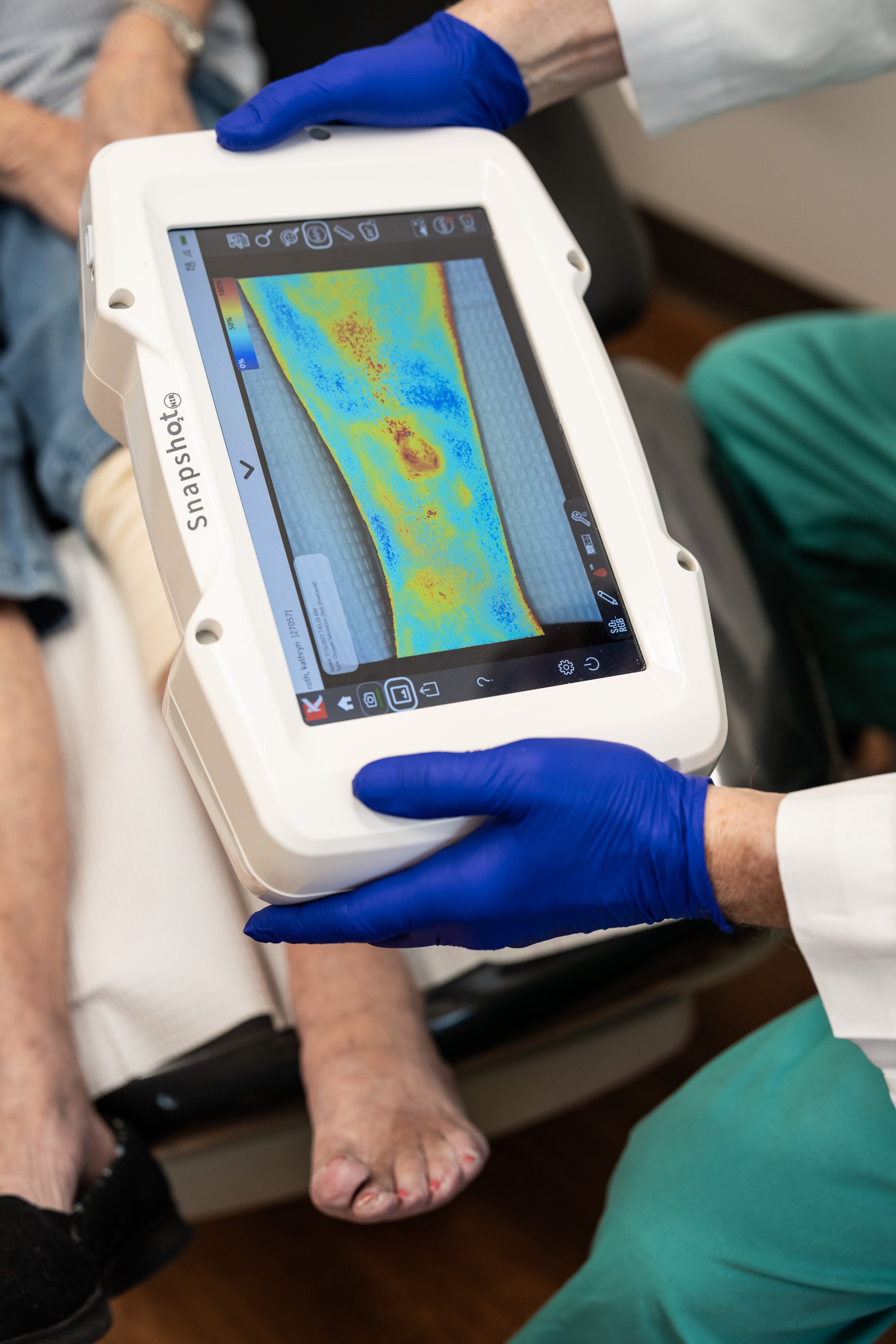
Tissue oxygenation image (StO2) with Hemoglobin view on SnapshotNIR showing healing progress on the patients wound.
THE POWER OF PICTURES
Of course, it’s not just the LEAP patients at Gulf South Foot & Ankle who undergo imaging with Snapshot—practitioners at all three clinic locations use the device to assess and track wounds and vascular issues in non-diabetic patients as well. “It’s been incredibly helpful”, says Dr. Watkins, “not only in attaining patient buy-in when it comes to accepting a diagnosis and treatment plan but also as a means of improving patient morale throughout a treatment pathway.” That’s because the clinicians can demonstrate quite easily, through captured Snapshot images, when a wound is healing, or blood flow is improving.
In Kathryn Roth’s case, Dr. Watkins is still using Snapshot to track the healing progress of that small ulcer on her leg. “The last time I saw her, the ulcer had calmed down quite a bit,” he says, adding that Roth is still being treated with intravenous antibiotics, but on an outpatient basis.
For her part, Roth, who has been a patient at Gulf South Foot & Ankle for more than 10 years, says she’s grateful that Dr. Watkins was able to capture Snapshot images that clearly demonstrated her need for medical intervention. “Thanks to that device, Dr. Watkins was the first one to notice the infection in my leg,” she says. “But that’s no surprise. He’s really good. He’ll do anything for his patients.”
GAME CHANGERS: If you would like to share your experience with SnapshotNIR through an Ask the Expert Interview or Customer Story, contact Kent Imaging via email for more details or call TF: 1-833-733-5368