
Addressing Common Difficulties in the Wound Care Setting
Time is tissue, yet effective wound assessment can be a real challenge.
Common barriers and difficulties in wound care settings include:
Assessing and treating underlying conditions to chronic wounds
Inaccurate wound measurements and documentation of wound size
Lack of non-invasive methods to assess wounds
Lack of consistency between assessment methods
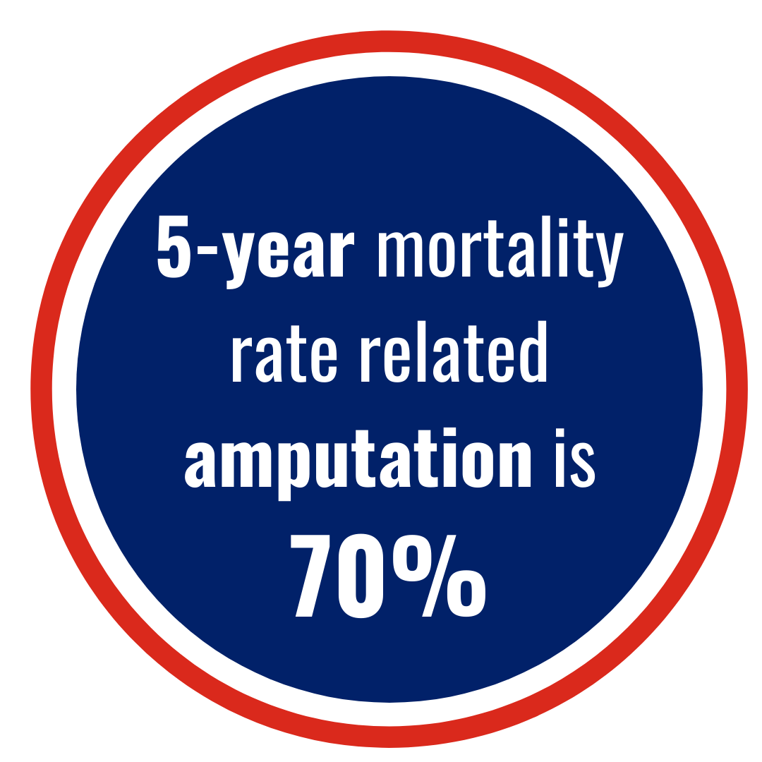
Chronic non-healing wounds affect millions of people every year. These wounds can have a significant effect on a patient’s quality of life, with an increased risk of serious complications. Any open wound is a risk for infection, hospitalization, amputation, and even death. Wounds occur secondary to the disease process and are not considered actual diseases. Healthcare providers assess wounds and develop care plans based on etiology.
Some specific wound healing complications can include infection, tissue necrosis, osteomyelitis, peri-wound dermatitis, edema, and dehiscence. To add to this, common challenges in wound management processes can be inaccurate wound measurement, inaccurate or incomplete documentation, time-consuming workflows, lack of direct oversight to enforce wound care protocols, the inability to accurately assess patients non-invasively, and in some cases a lack of wound expertise.
Available assessment methods that are time-consuming, costly and inconsistent:
Ankle-brachial index (ABIs)
29% of Critical Limb Ischemia (CLI) patients have near normal ABIs, yet clinicians make decisions every day based on ABIs (1)
Transcutaneous oximetry (TCOM)
With the inability to place electrodes on the wound, this method can only assess the peri-wound
Considering the diagnostic utility of ABI testing and other forms of traditional assessment, how can clinicians better understand what is going on underneath the surface?
Is there a better way to assess and monitor wound healing progression?
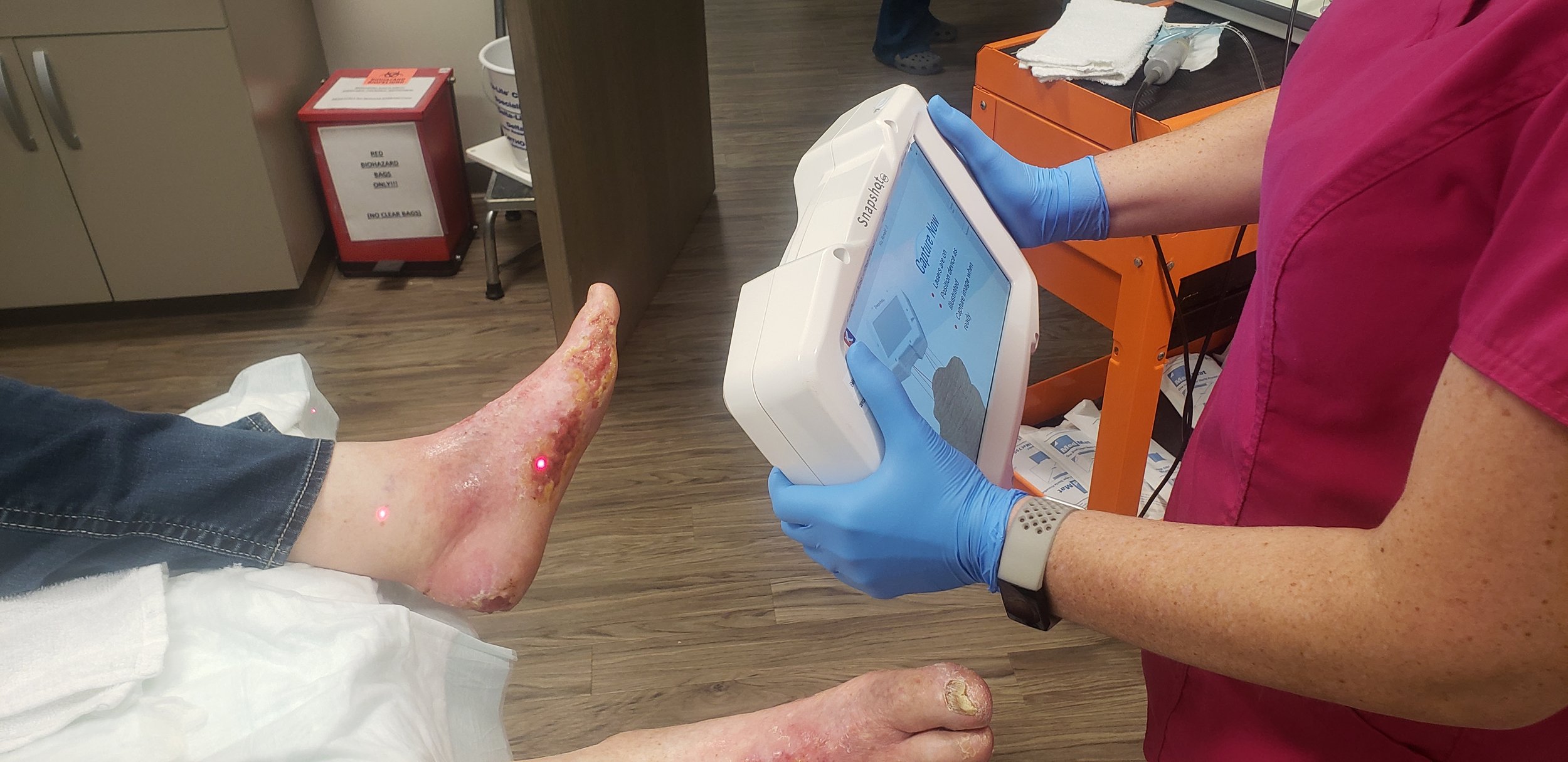
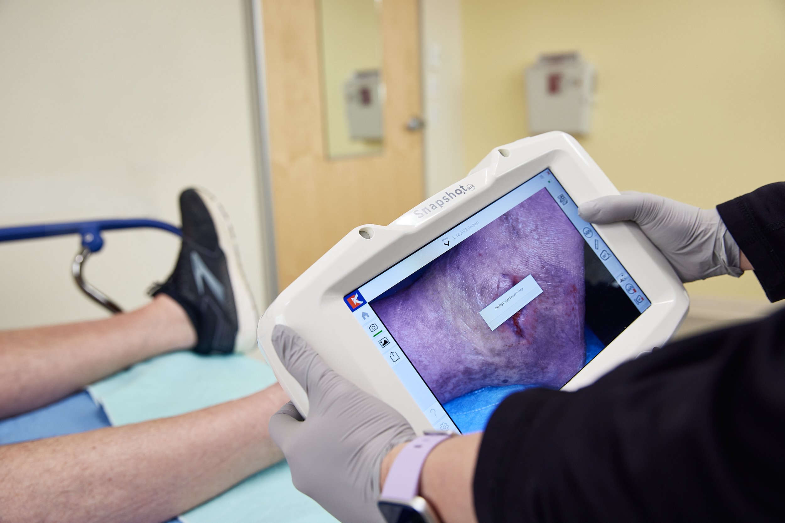
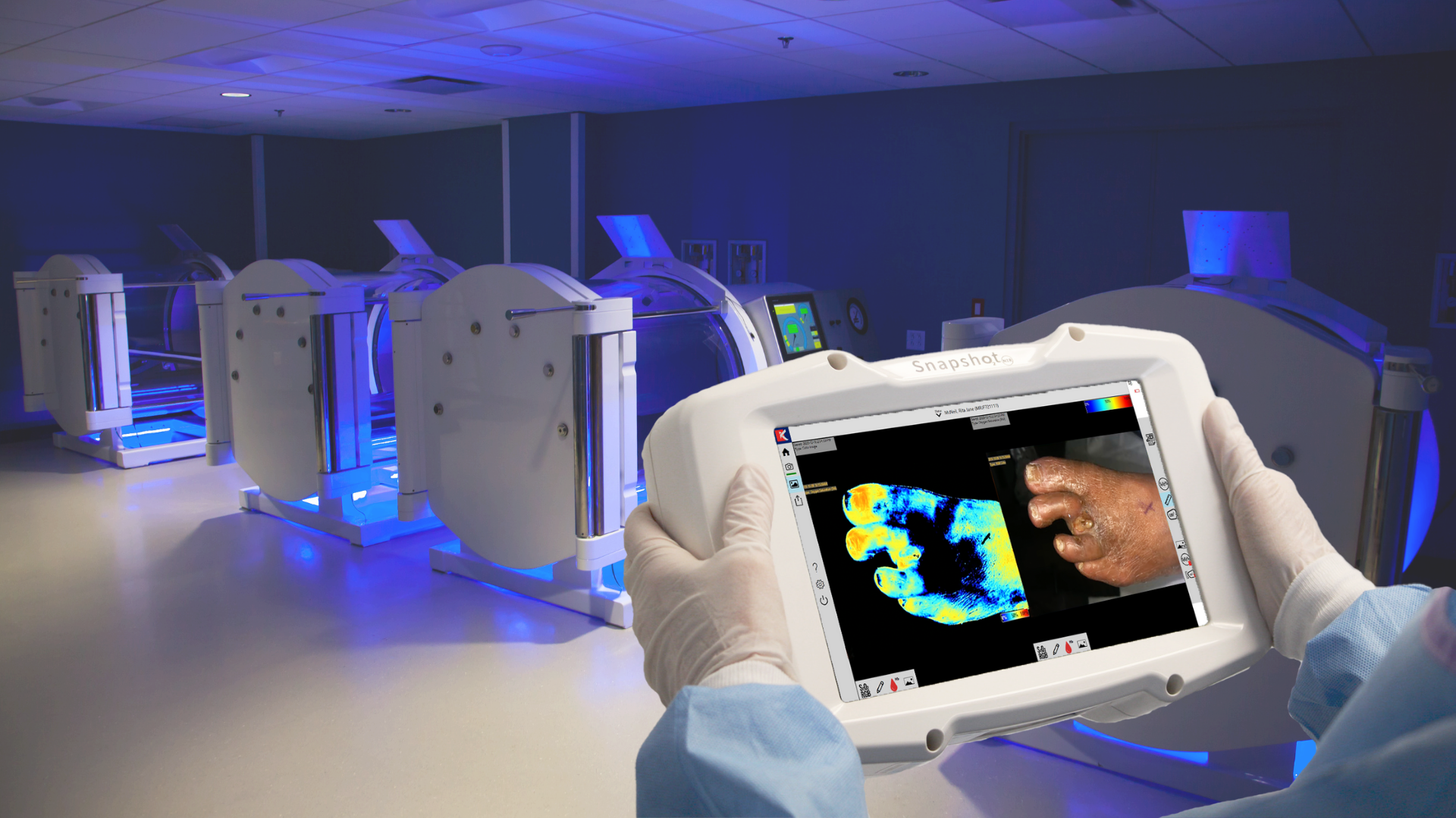
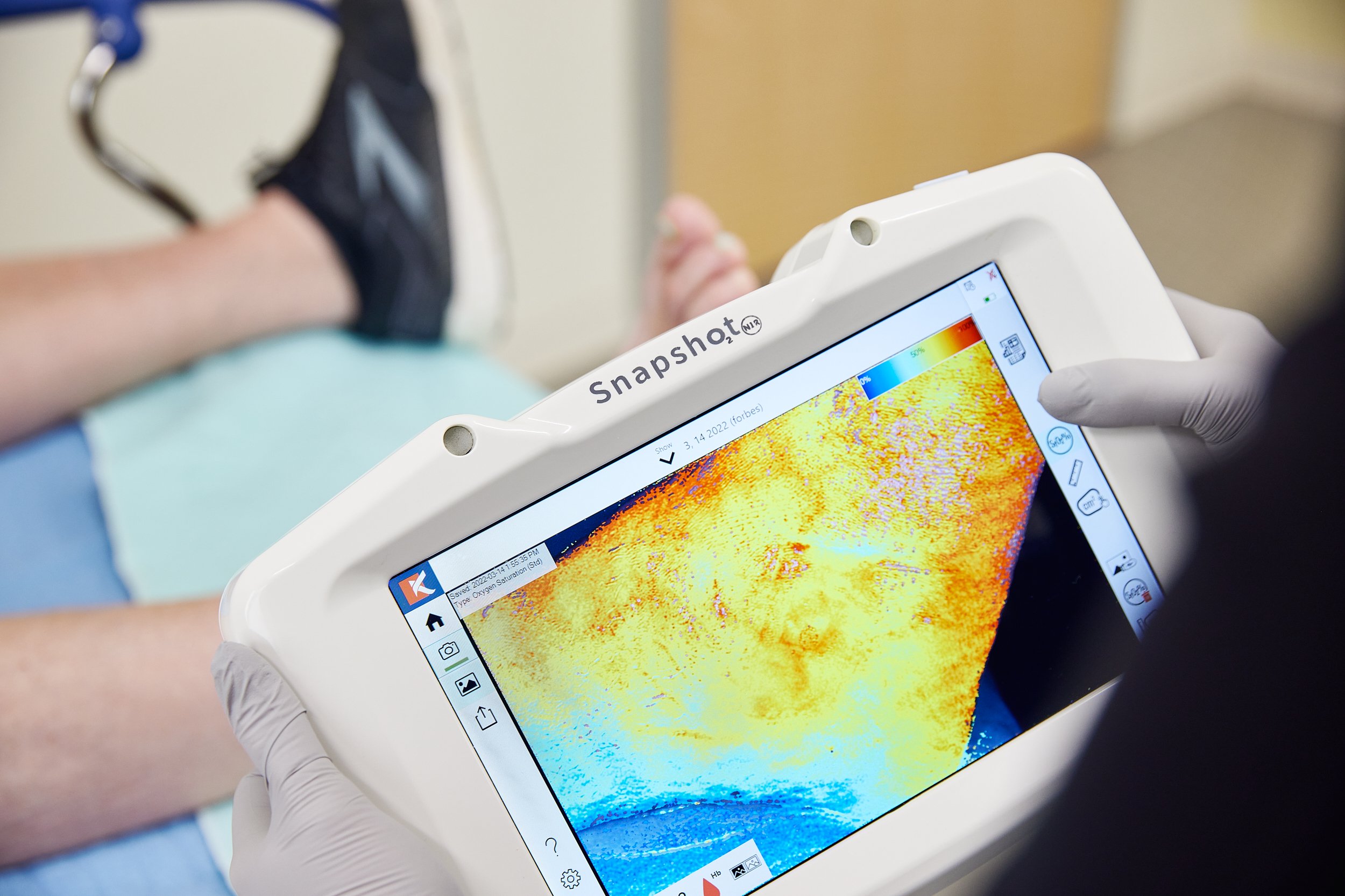
Using near-infrared spectroscopy imaging with SnapshotNIR, clinicians can gain near-instantaneous insight into “what lies beneath,” through the capture and display of tissue oxygenation in and around a wound. SnapshotNIR is a diagnostic-driven tool that can help overcome many of these wound care challenges by providing critical data to clinicians at the point of care. The device images may affect the plan of care by reducing decision-making time and potentially creating a more effective plan. If a treatment path is not providing the expected benefits, SnapshotNIR can support and document the decision to implement a change more quickly to expedite wound healing.
SnapshotNIR is lightweight, handheld, and portable, allowing users to capture tissue oxygenation images in just seconds with a click of a button with zero patient contact. Clinicians can triage patients earlier in the care process and expedite referral to integrated specialties, including vascular, enhancing interdisciplinary communication.
SnapshotNIR provides the ability to capture linear and area measurements directly on the images and store these in the patient record for easy numerical and visual comparison over time, speeding-up clinical workflow.
Additionally, the hemoglobin view (Hgb View) is a unique feature of SnapshotNIR’s imaging technology. Hgb View allows the clinician to infer tissue oxygenation as well as tissue perfusion. Tissue oxygenation provides insight into the metabolic health of the tissue showing how well the oxygen needs of the tissue are being met. The overall value of using hemoglobin view is that it provides clinicians and health care professionals with additional metrics, above and beyond the standard tissue oxygen saturation view.
The ease of use and ability to capture multiple modes of data can help improve patient outcomes and reduce healthcare costs.
Key Features of SnapshotNIR:
Point of Care Diagnostics
Non-Contact and Non-invasive
Captures oxyhemoglobin, deoxyhemoglobin, total hemoglobin, (StO2), & clinical (RGB) images
Capture linear and circumferential wound measurement
Immediately identifies ischemic tissue and the need for vascular intervention
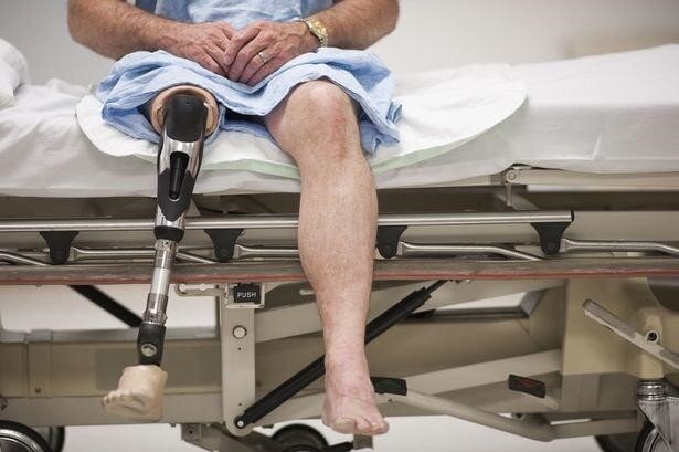
Medical decision-making through tissue assessment to prevent or identify the need for amputation
Comparable imaging to validate the therapeutic effect or change in care plan
Export images to EMR
Customized reports documenting outcomes - choose what type of image reports you want; a single image set or a two-image comparison
WiFi connectivity - quickly set up to email image reports
To review a breadth of case studies of Kent’s near-infrared spectroscopy imaging device, SnapshotNIR, go here.
To review physician opinions and experiences on using SnapshotNIR in the modern wound care clinic, explore our education page, which features interviews, webinars, and customer stories.
Source: https://www.woundsource.com/blog/complications-in-chronic-wound-healing-and-associated-interventions
References
(1) Heterogeneity of Ankle-Brachial Indices in Patients Undergoing Revascularization for Critical Limb Ischemia Devraj Sukul, MD,a Scott F. Grey, PhD,a Peter K. Henke, MD,b,c Hitinder S. Gurm, MD,a,c and P. Michael Grossman, MDa,c JACC Cardiovasc Interv. 2017 Nov 27; 10(22): 2307–2316.