
What is the difference between Thermography and NIRS?
There is a distinct difference between thermography and near-infrared spectroscopy.
The technologies differ in what they measure and what light is captured to form the images.
SnapshotNIR uses reflected near-infrared (NIR) light (600nm-1000nm) and measures oxygenated and deoxygenated hemoglobin.
Thermography reports the temperature of tissue by measuring the infrared light (longer wavelengths) emitted at the surface of the tissue.
There can be a correlation between temperature and blood flow, but SnapshotNIR measures more than flow and is thus largely independent of temperature. The StO2 image generated by SnapshotNIR shows the balance between hemoglobin oxygen delivery and oxygen consumption in superficial tissue. The clinician can use these diagnostic-driven images to help assess whether the oxygen supply is meeting demand. For example, when using SnapshotNIR in surgery you can look for changes and trends in oxygenation. At risk tissue can be determined by trending lower oxygenation numbers over the course of the procedure.
Thermography or thermal imaging is a process where a thermal camera captures and creates an image of an object by using infrared radiation emitted from an object. The amount of radiation emitted by an object increases with temperature; therefore, visualizing temperature variations. NIRS detects unique 'fingerprints' in the electromagnetic spectrum. Known as spectral signatures, these 'fingerprints' enable the identification of the materials that make up a scanned object. For example, a spectral signature for oxygenated hemoglobin visualizes the level of tissue oxygenation guiding clinicians in assessing disease. So, thermography visualizes an object's temperature, while NIRS visualizes an object's chemical nature.
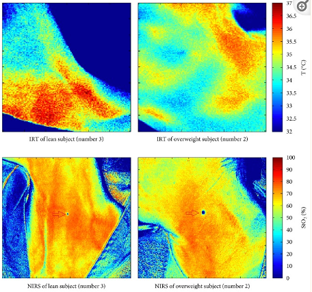
J Healthc Eng. 2017; 2017: 5986452. Published online 2017 Sep 14. doi: 10.1155/2017/5986452 Multimodal Imaging for the Detection of Brown Adipose Tissue Activation in Women: A Pilot Study Using NIRS and Infrared Thermography
Valentina Hartwig, 1 Letizia Guiducci, 1 Martina Marinelli, 1 Laura Pistoia, 2 Tommaso Minutoli Tegrimi, 2 Giorgio Iervasi, 1 Alfredo Quinones-Galvan, 2 and Antonio L'Abbate 1 , 3
“Thermography visualizes an object’s temperature, while NIRS visualizes an object’s chemical nature.”
Case Study
In a recent case study, Dr. Glyn Jones presents a patient who underwent bilateral skin-sparing mastectomies (the left breast mastectomy was prophylactic) with immediate pre-pectoral direct-to-implant (DTI) reconstruction with Natrelle SSF 450mL SoftTouch gel implants and AlloDerm. Thermography and tissue oxygen saturation (StO2) images were captured at five time points: pre-mastectomy, post-mastectomy, immediate post-reconstruction, 1-hour post-reconstruction, and 1-day post-reconstruction.
StO2 images captured through near-infrared spectroscopy (SnapshotNIR) showed the flaps to be well-oxygenated and viable, whereas the FLIR thermography images showed an area of potential concern. SnapshotNIR matched clinical judgment and the surgeon chose to proceed immediately with the planned DTI procedure. The mastectomy flap survived postoperatively and completed healing without complication.
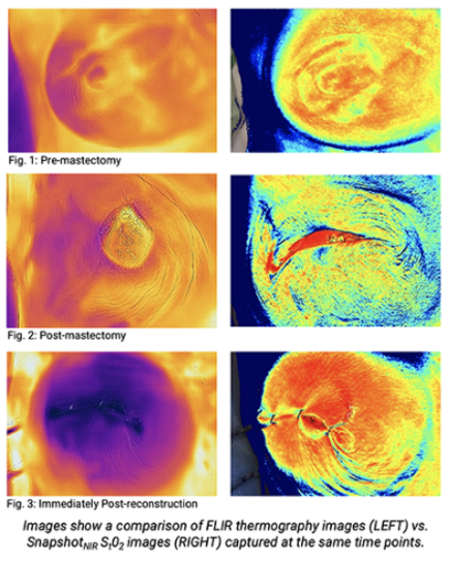

Review the full case study, Comparing StO2 and Thermography Images in Bilateral Skin-sparing Mastectomy, from Dr. Glyn Jones, MD.
To speak to a product expert about additional resources or to schedule a demo, go here.
To stay on top of Kent news, sign up for our quarterly newsletter here which features the latest clinical publications, case studies, customer stories, expert interviews, and more.
References:
First image study: J Healthc Eng. 2017; 2017: 5986452. Published online 2017 Sep 14. doi: 10.1155/2017/5986452 Multimodal Imaging for the Detection of Brown Adipose Tissue Activation in Women: A Pilot Study Using NIRS and Infrared Thermography Valentina Hartwig, 1 Letizia Guiducci, 1 Martina Marinelli, 1 Laura Pistoia, 2 Tommaso Minutoli Tegrimi, 2 Giorgio Iervasi, 1 Alfredo Quinones-Galvan, 2 and Antonio L'Abbate 1, 3
Second image study: case study by Dr. Glyn Jones, Comparing StO2 and Thermography Images in Bilateral Skin-sparing Mastectomy