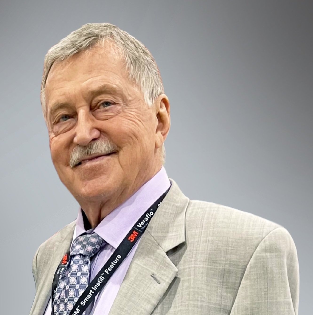
ASK THE EXPERT: DR. CHARLES ANDERSEN
ASK THE EXPERT
Dr. Charles Andersen is the Chief of Vascular/Endovascular/Limb Preservation services (Emeritus) Chief of Wound Care Service, Madigan Army Medical Center, Tacoma, WA; Clinical Professor of Surgery, UW, USUHS.
KI: You recently published a poster where you looked at redefining wound healing, particularly in diabetic foot and venous ulcers. Tell us about the benefit you observed in using near-infrared spectroscopy imaging with SnapshotNIR to assess healing in DFU’s, venous ulcers, and other various wounds, and how this impacted patient outcomes.
CA: Traditionally, wound healing has been defined as the epithelialization of a wound. It’s been observed by many providers that using epithelialization as the endpoint to progress to a lower level of care may be prematurely halting offloading in the diabetic foot ulcer or stopping active compression in a venous leg ulcer.
Venous leg ulcers and diabetic foot ulcers have a high incidence of recurrence after healing. Due to this trend in reoccurrence, many clinicians have independently decided to extend the time that they continue to use a total contact cast or extend the time that a wrap is used on a venous leg ulcer.
Unfortunately, in the past, there has been no way to quantify the likelihood of reoccurrence, or if a wound is truly healed below the surface. In our clinic, we are now using SnapshotNIR to help decide when a wound is truly healed. Near-infrared spectroscopy imaging allows you to see the inflammation that remains underneath an epithelialized wound, indicating that deep tissue healing is still ongoing below the epithelialization. Using Snapshot, the clinician can continuously track and monitor the tissue healing – looking for normalized tissue oxygenation values in the surrounding wound tissues. When the oxygenation level below the wound is the same as the surrounding tissue, it indicates deep tissue healing. Deep healing underneath the epithelialized wound may take two or three weeks to complete.
Thanks to this serial imaging, we know that the wound is not only healed on the surface but healed below the surface as well. This insight can be used as an endpoint for wound healing. With this accurate and actionable data, we have noted a lower rate of recurrence. We can more definitively decide to progress to orthotics from the total contact cast or replace active compression with a two or three-layer wrap on a venous leg ulcer with a support stocking.
SnapshotNIR offers a truly different way to look at wound healing. We consider it a highly valuable tool in redefining wound care. I personally think that Snapshot very likely will become a standard in research for defining wound healing.
KI: How do you use SnapshotNIR on a typical wound care clinic day?
CA: Through the regular use of near-infrared spectroscopy (NIRS) imaging in the Wound Care Clinic at the Madigan Army Medical Center, we have concluded that the oxygenation images captured with Snapshot are very valuable in providing additional data and insight to evaluate the treatment plan. This data may be used to change the treatment plan or confirm the continuation of the current course of action. This data also supports the tracking and documentation of the wound healing progress. As a result, we have recently decided that each patient who visits the clinic will get an NIRS study as part of their evaluation.
Below is a review of just a few of the patients we saw at the clinic over a 2-day span in April 2022, which serves to illustrate the type of valuable information we can get from the routine use of NIRS imaging with the Snapshot device.
A patient presented with a blood-filled blister on the tip of the right hallux and a wound on the adjacent toe. The question asked on evaluation was “Is this vascular etiology, for example, a distal ischemia or blue-toe syndrome, or strictly trauma, with the ability to heal both wounds?” The NIRS study showed adequate perfusion, so no additional vascular studies were felt to be indicated. Local wound care was continued based on the demonstration of adequate oxygenation.
A female patient with a very large thoracic spine pressure injury was being treated at the wound care clinic due to the location of this pressure injury right down to the spine. She had undergone wound bed preparation and was treated with a wound VAC and an advancement flap. During the course of this treatment, NIRS was used on multiple occasions to assure adequate oxygenation of the flap. In the follow-up visit, the flap continued to be well perfused. There was an area of some erythema at the lateral margin of the advancement flap. Using both fluorescence imaging (MolecuLight) to detect bacterial load and NIRS imaging (Kent Imaging) to measure tissue oxygenation, we were able to determine that the area of erythema was inflammatory. There was no evidence of bacteria and the tissue had adequate oxygenation in the wound area to continue local wound care, with the anticipation that this part of the wound would continue to heal satisfactorily.
A fairly common problem seen in the wound clinic is moisture-related skin damage in the area of the buttocks or trochanteric region. The question becomes “How much damage is associated with the moisture-related skin damage?” This particular patient had some discoloration adjacent to the moisture-related skin damage which could possibly be related to a deep tissue injury. With evaluation using NIRS there was no significant inflammation in the area, ruling out any significant deep tissue injury. The discoloration was recognized to be just pigmentation and not tissue damage.
These examples and many others illustrate that SnapshotNIR can be very helpful in identifying early pressure injuries as well as in grading the degree of pressure or tissue damage in pressure injuries.
A patient with bilateral mastectomies for breast cancer with irradiated skin had developed a problematic chronic wound due to this irradiated skin tissue. She was treated with topical oxygen therapy with subsequent re-epithelialization of the wound. Throughout the process, she was monitored with NIRS. The NIRS documented increased tissue oxygenation as a result of the topical oxygen therapy. On this particular visit, the patient was fitted for a bra with prosthetic breasts. The NIRS imaging was used to identify that although the wound was epithelialized, there was an area of continued inflammation within the wound, located in an area where the bra would cross the wound. With the recognition that the wound was not fully healed and would perhaps be subject to breakdown with the pressure of the bra band, extra padding and protection were provided to that section of the wound to facilitate the wearing of the bra without tissue breakdown.
The next patient is a 51-year-old female with a history of chronic hidradenitis suppurativa in her right axillary area. To treat this, she had a wide incision in her axillary skin and subcutaneous tissue which contains the apocrine sweat gland. As a result, she had a large tissue defect and was being prepared for a split-thickness skin graft. The SnapshotNIR study showed uniform oxygenation of the granulation tissue. This is important information when proceeding with a split-thickness skin graft. The two things that we will now make routine prior to applying a split-thickness graft are (1) making sure there is no bacteria in the wound bed, evaluating and documenting with fluorescence imaging for bacteria, and (2) ensuring that the granulation tissue has uniform oxygenation (NIRS). As a result of these two evaluations, the patient is being prepared to proceed with the split-thickness skin graft.
An example of the extremely significant data that can be obtained with Snapshot is illustrated in this next patient - a female with severe bilateral arterial insufficiency, and a previous right below-knee (BK) amputation, developed progression of her arterial disease with a breakdown of her BK amputation and now has a large ischemic wound in this BK amputation. The patient was being prepared for a right above knee (AK) amputation. On this clinic day in advance of the surgery, the NIRS study documented not only very severe ischemia in the area of the BK wound, but it also indicated very poor oxygenation in the thigh. This was felt to be inadequate oxygenation to support an AK amputation. The recognition of this with the data from the NIRS study led to the scheduling of an additional vascular procedure: a right common femoral endarterectomy to help establish adequate perfusion for the patient to support the healing of the AK amputation. This was a critical finding - had this issue not been recognized in advance and the amputation proceed as originally scheduled, the AK amputation would likely have failed, leading to a very large wound and potentially, a very poor outcome.
Review a detailed case study conducted by Dr. Charles Andersen, MD.
GAME CHANGERS: If you would like to share your experience with SnapshotNIR through an Ask the Expert Interview, fill out this form.

